Various Organs Abnormalities

It has been shown that exposure to EMFs increases arrhythmias, heart attacks (myocardial infarctions), COPD, asthma, diabetes, autonomic imbalance, impaired liver and kidney function, and cataracts.
In recent years, arrhythmia, COPD, asthma, diabetes, liver disease, kidney disease, and cataracts are on the rise, and EMFs are likely to be one of the factors in their increase.
Recent Trends
Arrhythmias :
US
Japan
Heart Failure :
US
Japan
COPD :
US
Japan
Asthma :
US
Japan
Diabetes :
US
Japan
Liver Disease :
US
Japan
Impaired Liver Function :
Japan
Chronic Kidney Disease :
US
Japan
Acute Kidney Failure :
US
Cataracts :
UK
Table of ContentsAll_Pages
Arrhythmias and Heart Attacks
First, I will present studies showing that arrhythmias, heart attacks (myocardial infarctions), and other heart problems increased with exposure to EMFs from workplaces.

Among the arrhythmias, an increase in bradycardia was observed in particular. Bradycardia is a kind of arrhythmia where the heart rate is abnormally slow, less than 60 beats per minute.
Studies
Savitz et al. 1999
For male employees of five U.S. large power companies, deaths from arrhythmias and heart attacks (acute myocardial infarctions) increased as cumulative exposure to ELF-EMFs at workplaces increased.
This was especially more pronounced when looking at cumulative exposure prior to the past 20 years.
Increase in Arrhythmias
Increase in Heart Attacks
Tikhonova 2003
For personnel of civil aircraft radar tracking system in Russia, cardiovascular diseases increased when they were occupationally exposed to RF-EMFs, particularly millimeter waves.
This was especially more pronounced among younger people aged 30-39 years.
Increase in
Cardiovascular Diseases
Among the cardiovascular diseases, increases in arterial hypertension and ischemic heart diseases were observed. Heart attacks (myocardial infarctions) are well-known typical ischemic heart diseases.
Increase in Arterial Hypertension
The odds of arterial hypertension increased 2-fold with the millimeter wave exposure.
Increase in Ischemic Heart Diseases
The odds of ischemic heart diseases increased 8-fold with the millimeter wave exposure.
Sahl 2002
For male employees of a power company in California, deaths from heart atttacks (acute myocardial infarctions) increased when cumulative exposure to ELF-EMFs at workplaces was higher.
This was especially more pronounced when looking at cumulative exposure in the recent five years.
An increase in COPD was also observed, which is described in the section on COPD and Asthma.
Increase in Heart Attacks
Hakansson 2003
For adults on the Swedish twin registry, deaths from arrhythmias increased when ELF-EMFs at workplaces were stronger.
This was especially more pronounced among relatively young people aged 65 years or younger.
Also, somewhat of an increase in deaths from heart attacks (acute myocardial infarctions) was observed.
Increase in Arrhythmias
Increase in Heart Attacks
The mortality risk of heart attacks (acute myocardial infarctions) increased by 30% with 0.3 μT or more of the ELF-EMFs at the workplaces.
Wilén et al. 2003
For employees in nine Swedish factories, heart rates decreased and bradycardia increased when they used industrial machines that emit RF-EMFs, called RF Sealers.
RF Sealers are used to seal for tarpaulins, tents, rain clothes, and covers, and users were exposed to fairly strong RF-EMFs with an average strength of 2.6 mW/cm2.
Decrease in Heart Rates
The heart rate decreased by 10% with the use of RF Sealer.
Increase in Bradycardia
The number of episodes of bradycardia increased by 60% with the use of RF Sealer.
Also, an increase in electromagnetic hypersensitivity (EHS) was observed.
Increase in EHS
Déoux et al. 1997
This is a case report of a 47-year-old woman from France.
The woman worked in an office directly above an electrical transformer and was exposed to ELF-EMFs with an average strength of 23 μT for 40 hours per week for 22 months.
During the work period, a slow heart rate and bradycardia were observed in the woman.
After termination of the exposure, the heart rate recovered but did not return to its original level.
Increase in Bradycardia
The heart rate was drastically decreased and bradycardia occured with electrical transformer EMF exposure during work.
Since the heart rate did not return to its original level, it is possible that some irreversible damage had been done to the heart.
Other observations included frequent premature ventricular contraction. Frequent premature ventricular contraction can lead to fatal arrhythmias.
Kilicalp et al. 2009
Cell phones with a local SAR of 0.95 W/kg were placed under the floor of the breeding cages and kept on communication for 20 minutes per day, and guinea pigs were exposed to their EMFs for 30 days.
As a result, heart rates decreased and bradycardia resulted.
Also, treatment with green tea, which has antioxidant properties, suppressed the damage above.
Decrease in Heart Rates
The heart rates decreased by 30% and bradycardia resulted due to the cell phone EMF exposure. Green tea, which has antioxidant properties, suppressed it.
Also, prolongation of PR interval, a diagnostic criterion for atrioventricular block, was observed.
Prolongation of PR Interval
The PR interval increased 3-fold due to the cell phone EMF exposure. Treatment with green tea had no effect.
Damage to the Heart
Next, I will present studies showing that exposure to EMFs damaged the sinus node and heart muscle of the hearts of rats.

Damage to the Sinus Node
The loss of functional cells such as heart muscle cells and their replacement by structural component such as collagen is called fibrosis (scarring). (Piek et al. 2016)
Heart fibrosis results in stiffening and remodeling (structual and functional change) of the heart, and is increasingly recognized as a major cause of death in many chronic heart diseases. (Piek et al. 2016)
For example, fibrosis is an important risk factor for arrhythmias. (Kazbanov et al. 2016)
Bradycardia, an arrhythmia where the heart rate is abnormally slow, results from conduction block in the sinus node or atrioventricular node, which is often caused by fibrosis. (Verheule and Schotten 2021)
The sinus node is the heart muscle that produces the heart rhythm and is known as the natural pacemaker.
And, it has been shown that EMF exposure increases cell death in the heart muscle of rats and fibrosis in the sinus node.
Studies
Liu et al. 2015
Male rats aged 8 weeks, equivalent to adolescents, were exposed to pulse-modulated 2.9 GHz RF-EMFs with a strength of 50 mW/cm2 for 6 minutes per day for 12 months.
As a result, the sinus node of the heart was damaged and became fibrotic (scarred).
Damage to the Sinus Node
(short term)

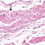

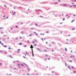
Due to the several weeks of EMF exposure, the sinus node was damaged.
Damage to the Sinus Node
(long-term)


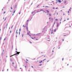
Due to the several months of EMF exposure, the sinus node became fibrotic (scarred). Also, decreased parenchyma cells (functinal cells) of the sinus node and infiltration of lipid droplets were observed.
Accumulation of lipid dropletsis is frequently observed in infectious, tumorous and other inflammatory conditions. (Bozza and Viola 2010)
Damage to the Sinus Node
(magnified)


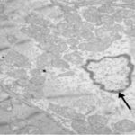

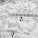
Due to the several weeks of EMF exposure, myofibrils (components of a muscle fiber) and mitochondria in the sinus node were damaged.
Fibrosis of the Sinus Node
The collagen content in the sinus node increased 4-fold due to the EMF exposure. This means fibrosis (scarring) of the sinus node.
Kiray et al. 2012
Adult male rats were exposed to 50 Hz ELF-EMFs with a strength of 3 mT for 4 hours per day for 2 months.
As a result, reactive oxygen species (ROS) increased in the heart, the heart muscle was damaged, and cell death in the heart muscle increased.
Increase in ROS
An increase in lipid peroxidation and a decrease in antioxidant activity mean an increase in ROS.
Damage to the Heart Muscle
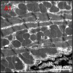
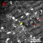
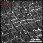
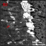
Due to the ELF-EMF exposure, myofibrils (components of a muscle fiber) and mitochondria in the heart muscle were damaged.
Increase in Cell Death 1
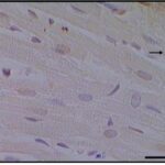
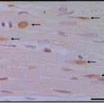
Due to the ELF-EMF exposure, cell death in the heart muscle increased.
Increase in Cell Death 2
Türedi et al. 2014
Pregnant rats were exposed to 900 MHz RF-EMFs with a strength of 50 μW/cm2 for 1 hour per day for 1 week in late pregnancy.
As a result, in the hearts of the born-son rats, the heart muscle was damaged, and cell death in the heart muscle increased.
Damage to the Heart Muscle
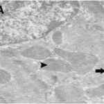
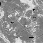
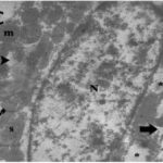
Due to the RF-EMF exposure, myofibrils (components of a muscle fiber) and mitochondria in the heart muscle were damaged.
Increase in Cell Death 1


Due to the RF-EMF exposure, cell death in the heart muscle increased.
Increase in Cell Death 2
The percentage of cell death in the heart muscle increased 3-fold due to the RF-EMF exposure.
COPD and Asthma
Next, I will present studies showing that chronic obstructive pulmonary disease (COPD) and interstitial pneumonia increased with exposure of men to EMFs from workplaces.

I will also present a study showing that asthma increased in children with exposure of pregnant women to EMFs.

Studies
Sahl 2002
For male employees of a power company in California, deaths from COPD increased when cumulative exposure to ELF-EMFs at workplaces was higher.
An increase in heart atttacks (acute myocardial infarctions) was also observed, which is described in the section on Arrhythmias and Heart Attacks.
Increase in COPD
The mortality rate of COPD increased 3-fold in the top 30% for cumulative exposure to the ELF-EMFs at the workplaces.
Milham 1985
For men aged 20 years or older in Washington, deaths from COPD and interstitial pneumonia increased when they were occupationally exposed to ELF-EMFs.
Also, increases in deaths from gastric ulcers, lymphoma, and leukemia were observed.
Increase in Lung Disease
The proportion of COPD among all causes of death incerased by 50% due to the occupational EMF exposure, and interstitial pneumonia by 40% .
Increase in Other Diseases
The proportion of lymphoma and leukemia among all causes of death incerased 2-fold due to the occupational EMF exposure.
Li 2011
For pregnant members of a major health maintenance organization (HMO), residing in the San Francisco Bay Area, asthma in children born to them increased as the daily average strength of ELF-EMFs they had been exposed to increased.
Increase in Asthma
The hazard of asthma in their children increased 1.7-fold with 0.03-0.2 μT of the ELF-EMF exposure of the pregnant women, and 3.5-fold with more than 0.2 μT.
The increase in the hazard of asthma means an increase in the incidence rate of asthma per unit of time (e.g., per year).
The following graph shows the incidence rate of asthma in children by age.
Increase in Asthma by Age
The incidence rate of asthma in children at age 12 increased 1.6-fold from 16% to 26% with 0.03-0.2 μT of the ELF-EMF exposure of the pregnant women, and 2.9-fold to 46% with more than 0.2 μT.
Booth et al. 2001
For residents aged 15-72 years in the Auckland metropolitan area, New Zealand, asthma increased when ELF-EMFs from high-voltage lines were stronger.
An increase in diabetes was also observed, which is described in the section on Diabetes.
Increase in Diabetes
The odds of chronic illnesses increased 2-fold, and asthma 3-fold in the top 40% for the strength of the ELF-EMF from the high-voltage lines.
Damage to the Lungs
Next, I will present a study showing that exposure to EMFs damaged the lungs of rats, and stunted their growth.

I will also present studies showing that exposure to EMFs increased reactive oxygen species (ROS) in the lungs of rats.

Stunted Lung Growth
COPD is conventionally thought of as a disease of adult smokers. However, there is important epidemiological evidence that early life events, including pre-birth influences on lung growth, program the child to be at increased risk for future COPD. (Bush 2008)
A study result suggested that airway function throughout adult life may be determined during fetal development and the first few months of post-birth life. (Gibson and Simpson 2009)
Also, a study found that children developing asthma by age 7 had a lung function deficit and increased airway sensitivity as newborns and that this lung function deficit progressed to age 7. (Bisgaard et al. 2012)
Studies
Yahyazadeh et al. 2021
Pregnant rats were exposed to 900 MHz RF-EMFs with a strength of 1 mW/cm2 for 3 weeks throughout the entire pregnancy.
As a result, the lungs were damaged and their growth was stunted in the born-son rats.
Also, treatment with melatonin, which has antioxidant properties, suppressed the damage above.
Damage to the Lungs
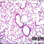
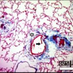
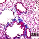
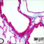
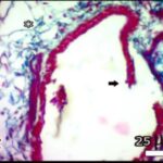
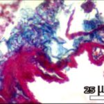
Due to EMF exposure during pregnancy, the bronchioles and connective tissue within the lungs of the son rats were damaged. Melatonin, which has antioxidant properties, suppressed it.
Stunted Lung Growth
Gharib 2011
Adult male rats were exposed to 950 MHz RF-EMFs for 1 hour per day for 50 days.
As a result, reactive oxygen species (ROS) increased in the lungs, heart, and blood, and lactate dehydrogenase (LDH), an organ damage marker, increased.
Also, treatment with kombucha, which has antioxidant properties, suppressed the damage above.
Increase in ROS in the Lungs
Increase in ROS in the Heart
Increase in ROS in Blood
The total antioxidant capacity decreased by 60% due to the RF-EMF exposure. Also, the kombucha, which has antioxidant properties, suppressed it.
An increase in lipid peroxidation and decreases in antioxidant activity and total antioxidant capacity mean an increase in ROS.
Organ Damage
The LDH (lactate dehydrogenase) activity, an organ damage marker, increased by 20% due to the RF-EMF exposure. Also, the kombucha, which has antioxidant properties, suppressed it.
Baltaci et al. 2012
Adult male rats were exposed to 50 Hz ELF-EMFs with a strength of 5 μT for 5 minutes per day, every other day, for 6 months.
As a result, reactive oxygen species (ROS) increased in the lungs and liver.
Also, treatment with zinc, which has antioxidant properties, suppressed the damage above.
Increase in ROS
An increase in lipid peroxidation mean an increase in ROS.
Diabetes
Next, I will present studies showing that diabetes increased with exposure to EMFs from homes and cell towers.

Studies
Booth et al. 2001
For residents aged 15-72 years in the Auckland metropolitan area, New Zealand, diabetes increased when ELF-EMFs from high-voltage lines were stronger.
An increase in asthma was also observed, which is described in the section on COPD and Asthma.
Increase in Diabetes
The odds of chronic illnesses increased 2-fold, and diabetes 7-fold in the top 40% for the strength of the ELF-EMF from the high-voltage lines.
Meo et al. 2015
For male students aged 12-17 years in two schools in Riyadh, Saudi Arabia, obesity increased and blood sugar and pre-diabetes somewhat increased when they attended the school with higher RF-EMFs from cell towers.
The strength of the RF-EMFs was 0.01 μW/cm2 in the stronger school, and 0.002 μW/cm2 in the weaker school.
It should be noted that the EMFs are not that strong in both schools, and the difference between the schools are small.
Increase in Obesity
The body mass index (BMI) increased by 1.4 percent points with attendance at the school with stronger EMFs from the cell towers.
Increase in Blood Sugar
The blood sugar level increased by 0.1 percent point with attendance at the school with stronger EMFs from the cell towers.
Increase in Pre-Diabetes
The percentage of pre-diabetes increased by 10% with attendance at the school with stronger EMFs from the cell towers.
Damage to the Pancreas
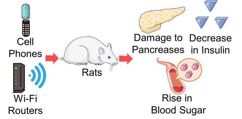
Next, I will present studies showing that exposure to EMFs from cell phones, Wi-Fi, and other sources damaged the islets of Langerhans in the pancreases of rats, decreased insulin, and raised blood sugar.
Damage to Islets of Langerhans
An organ deeply involved in diabetes is the pancreas. The two main roles of the pancreas are the secretion of digestive fluids called pancreatic juice and the secretion of hormones that regulate blood sugar levels.
Within the pancreas scattered clusters of cells that secrete hormones called islets of Langerhans, here and there.
Typical cells include alpha cells, which secrete glucagon, which raises blood sugar levels, and beta cells, which secrete insulin, which lowers blood sugar levels.
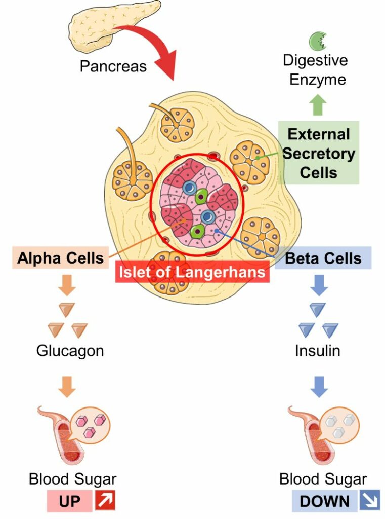
Langerhans cells are composed of about 70% beta cells and 20% alpha cells, with insulin-secreting beta cells accounting for the largest percentage.
In addition to glucagon, other hormones that raise blood sugar levels include cortisol and adrenaline, while insulin is the only hormone that lowers blood sugar levels.
Therefore in the pancreas, damage to the islets of Langerhans and a decrease in cell mass is likely to result in a decrease in insulin secretion, leading to a rise in blood sugar levels and, consequently, an increase in diabetes risk.

In fact, In type 1 diabetes, β-cell mass is reduced by as much as 70–80% at the time of diagnosis, In type 2, 25-50%. (Cnop et al. 2005)
And, it has been shown that exposure to EMF damages the islets of Langerhans in rats, decreases cell mass, decreases insulin secretion, and raises blood sugar levels.
Insulin Resistance
When blood sugar levels rise, the pancreas secretes insulin. Next, the liver, muscle, and fat cells receive insulin and begin to take up sugar (glucose) from the blood. This causes a fall in blood sugar levels.
On the other hand, at times, despite the rise in blood sugar and the subsequent insulin secretion, the liver, muscles, and fat cells will not take up sugar (glucose), and the blood sugar levels remain raised.
This is called insulin resistance.
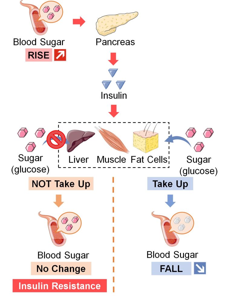
Insulin resistance is known to be common in patients with type II diabetes.
A study showed that EMF exposure increased insulin resistance in rats. One study alone is clearly insufficient evidence, but some corroboration is possible in terms of the following mechanism.
Mechanism of Insulin Resistance by EMFs
When muscle, liver, and fat cells receive insulin, a protein called GLUT4, which transports sugar (glucose), is activated and moves to the cell membrane.
Then, sugar (glucose) is taken up into the cell via this GLUT4.
However, when reactive oxygen species (ROS) increase, GLUT4 remains inactivated despite the insulin secretion, resulting in blood sugar levels remaining raised and insulin resistance. (Tsuruzoe et al. 2006, Mukherjee et al. 2013)
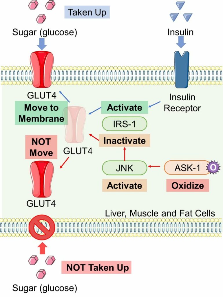
And numerous studies have shown that EMFs increase ROS.
Therefore, it is likely that when EMF exposure increases ROS, GLUT4, a transporter of sugar (glucose), becomes ineffective, resulting in insulin resistance.
Studies
Khaki et al. 2015
Rats aged 12 weeks, equivalent to adolescents, were exposed to 50Hz ELF-EMFs with a strenth of 3 mT for 4 hours per day for 6 weeks.
As a result, the islets of Langerhans within the pancreas shrank considerably, and blood insulin levels decreased.
Shrinkage of the Islet
of Langerhans 1
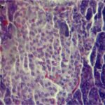
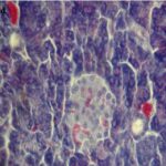
The islets of Langerhans shrank considerably due to the ELF-EMFs.
Shrinkage of the Islet
of Langerhans 2
Decrease in Blood Insulin Levels
The blood insulin level decreased by 40% due to the ELF-EMF exposure.
Mortazavi et al. 2016
Cell phones with a local SAR of 1.15 W/kg were placed and kept in talk mode for 6 hours per day, and adult female rats were exposed to their EMFs at a distance of 5 cm for 7 days.
As a result, the islets of Langerhans within the pancreas were damaged, and blood insulin levels decreased.
Damage to the Islet of Langerhans


Most cells in the islets of Langerhans were dissociated due to the cell phone EMF exposure.
Decrease in Blood Insulin Levels
The blood insulin level decreased by 50% due to the cell phone EMF exposure.
Bahaoddini et al. 2008
Male rats aged 12 weeks, equivalent to adolescents, were exposed to 50 Hz ELF-EMFs with a strength of 500 μT for 10 hours per day for 2 months.
As a result, blood insulin levels decreased, and blood sugar levels increased.
Increase in Blood Insulin Levels
The blood insulin level decreased by 40% due to the ELF-EMF exposure.
Increase in Blood Sugar Levels
The blood sugar level increased by 20% due to the ELF-EMF exposure.
Masoumi et al. 2018
A Wi-Fi router was placed and kept on data transfer for 4 hours per day, and male rats aged 12 weeks, equivalent to adolescents, were exposed to their EMFs at a distance of 30 cm for 45 days.
As a result, reactive oxygen species (ROS) increased in the pancreas, and the insulin secretory capacity of the islets of Langerhans was impaired.
Also, a glucose tolerance test showed a decrease in blood insulin levels and an increase in blood sugar levels.
Increase in ROS
An increase in lipid peroxidation and a decrease in antioxidant activity mean an increase in ROS.
Next are the results of the insulin secretory capacity of the islets of Langerhans.
This compared the amount of insulin secretion when the islets of Langerhans removed from the mice were exposed to low or high levels of sugar (glucose).
Impaired Insulin Secretory Capacity
The insulin secretory capacity of the islets of Langerhans was impaired due to the Wi-Fi EMF exposure. Exposure to low levels of sugar (glucose) decreased insulin secretion by 20% and to high levels by 50%.
Next are the results of the glucose tolerance test.
This compared the time course and the area under the curve (AUC) of blood insulin levels and blood sugar levels after injection of sugar (glucose) into the body cavity in fasting mice.
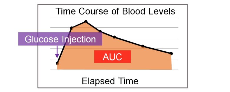
Decrease in Blood Insulin Levels
Increase in Blood Sugar Levels
The glucose tolerance test is used to determine if a person has diabetes.
Although there are no clear criteria in rats, there is no doubt that the Wi-Fi EMF exposure increased the risk of diabetes.
Meo and Rubeaan 2013
Cell phones were placed in the center of breeding cages and kept on incoming calls for 15-60 minutes per day, and male rats aged 60 days, equivalent to adolescents, were exposed to their EMFs for 3 months.
As a result, both fasting blood insulin levels and fasting blood sugar levels increased, resulting in an increase in insulin resistance index.
Increase in Blood Insulin Levels
The blood insulin levels increased by 20% for 30-45 minutes per day of the cell phone EMF exposure and by 10% for 45-60 minutes.
Increase in Blood Sugar Levels
The blood sugar levels increased by 60% for 30-45 minutes per day of the cell phone EMF exposure and by 70% for 45-60 minutes.
Increase in Insulin Resistance Index
The insulin resistance index increased by 90% for 30-45 minutes per day of the cell phone EMF exposure and by 80% for 45-60 minutes.
Autonomic Imbalance
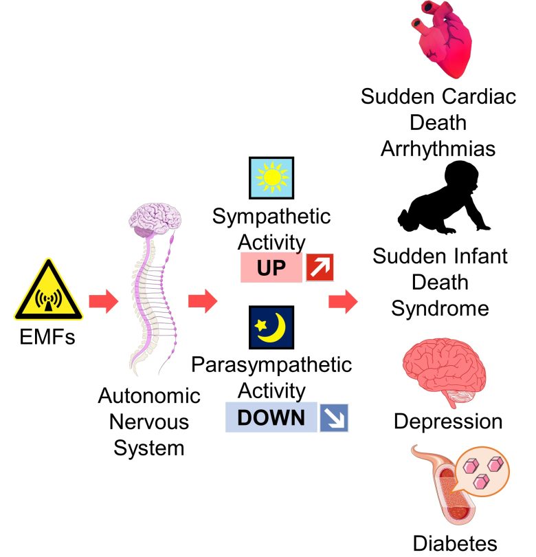
Next, I will present studies showing that among the autonomic nervous system, sympathetic activity increased and parasympathetic activity decreased with exposure to EMFs from high-voltage lines, radio towers, and cell phones.
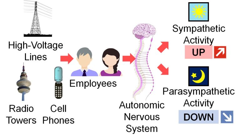
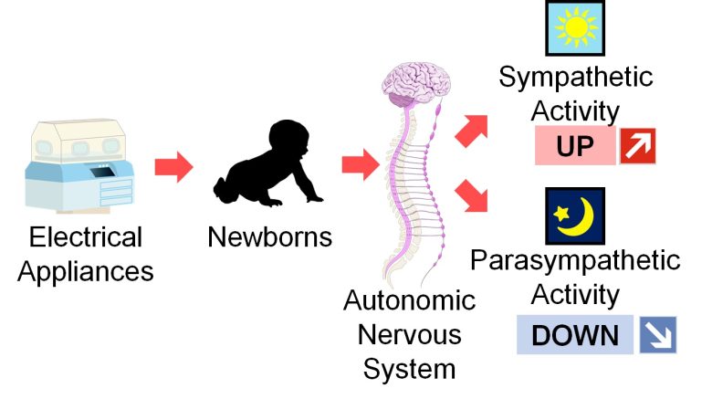
I will also present a study showing that sympathetic activity increased and parasympathetic activity decreased with exposure of newborns to EMFs from electrical appliances.
What is Autonomic Nerves ?
Autonomic nerves are peripheral nerves that extend from the brain and spinal cord and are distributed throughout the body, and they regulate body functions independently of our will.
There are mainly two types of autonomic nerves: sympathetic nerves, which are active during the daytime activities, and parasympathetic nerves, which are active during the nighttime rest.
The sympathetic and parasympathetic nerves each act on different organs of the body and have opposing effects on many organs.
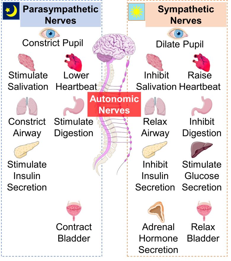
As described below, autonomic imbalance, particularly increased sympathetic activity and decreased parasympathetic activity, is correlated with various diseases.
Evaluation of Autonomic Nervous System by Heart Rate Variability
What is Heart Rate Variability ?
If you look at the activity of the heart over a period of time, such as with an electrocardiogram, you will see that the beat-to-beat interval fluctuates over time: 810 milliseconds, 830 milliseconds, 790 milliseconds, 835 milliseconds, and so on.
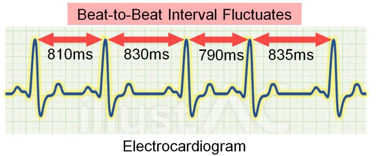
( Cited and Modified from Nyxo )
And this beat-to-beat interval fluctuates periodically, as shown in the graph below.
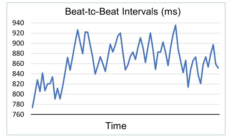
Heart Rate Variability
( Data taken from Ebrahimzadeh et al. 2014 )
This periodic variation in beat-to-beat intervals is called heart rate variability, and the autonomic nervous system can be evaluated by various parameters of heart rate variability.
Parameters of Heart Rate Variability (Time Domain)
The parameters of heart rate variability in the time domain include the following:
SDNN
It is the standard deviation of the beat-to-beat interval. In other words, it represents the amplitude of the variability of the beat-to-beat interval.
SDNN is the global marker, and reflects both sympathetic and parasympathetic modulation of heart rate. (Xhyheri et al. 2012)
Patients with SDNN values below 50 ms are classified as unhealthy, 50–100 ms have compromised health, and above 100 ms are healthy. (Shaffer and Ginsberg 2017)
RMSSD
It is the root mean square of the difference between adjacent beat-to-beat intervals.
It is a measure of parasympathetic activity, and parasympathetic activity is regarded as impaired when RMSSD is 25 ms or less. (Xhyheri et al. 2012)
PNN50
It is the percentage of adjacent beat-to-beat intervals with a difference of 50 ms or more.
It is a measure of parasympathetic activity, and parasympathetic activity is regarded as impaired when PNN50 is less than 3%. (Xhyheri et al. 2012)
Frequency Components of Heart Rate Variability
Heart rate variability consists of multiple frequency components, including those that fluctuate quickly, or at high frequency, and those that fluctuate slowly, or at low frequency.
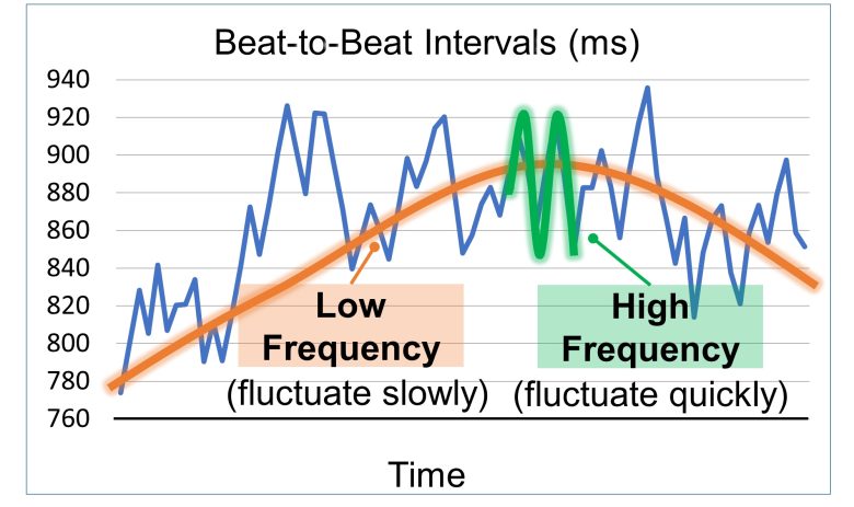
Heart rate variability consists of multiple frequency components
( Data taken from Ebrahimzadeh et al. 2014 )
The power by frequency of heart rate variability is illustrated below.
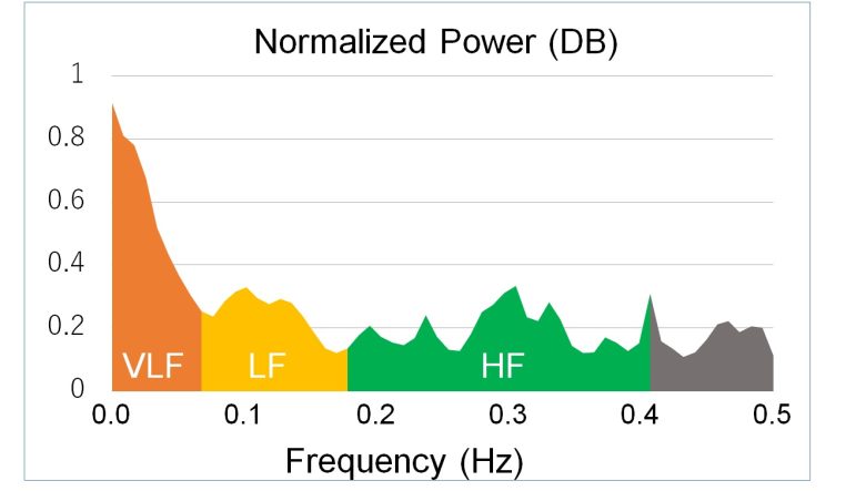
Power Spectral Density of Heart Rate Variability
( Data taken from Ebrahimzadeh et al. 2014 )
The parameters of heart rate variability in this frequency domain can also evaluate the autonomic nervous system.
Parameters of Heart Rate Variability (Frequency Domain)
The parameters of heart rate variability in the frequency domain include the following:
HF
It is the High Frequency range of 0.15 to 0.4 Hz, and a marker for parasympathetic activation. (Xhyheri et al. 2012)
LF
It is the Low Frequency range of 0.04 to 0.15 Hz and is considered a marker for sympathetic activation or for autonomic balance. (Xhyheri et al. 2012)
VLF
It is the Very Low Frequency range of 0.0033 to 0.04 Hz, and its relationship to the autonomic nervous system is currently unknown. (Xhyheri et al. 2012)
LF/HF Ratio
It is the index of the interaction between sympathetic and parasympathetic activity, and low indicates parasympathetic dominance and high indicates sympathetic dominance. (Xhyheri et al. 2012)
Decreased Heart Rate Variability and Autonomic Imbalance
Overall, low heart rate variability value usually indicate a relative sympathetic dominance, which may be due to high sympathetic activity and/or low parasympathetic activity. (Xhyheri et al. 2012)
Studies
Bortkiewicz et al. 2006
For service workers at 4 high-voltage substations and 4 radio link stations (*) in Poland, heart rate variability was altered when they worked at the high-voltage substations.
Workplace with no EMF exposure.
It showed increased sympathetic activity and relative sympathetic dominance.
Workers at the high-voltage substations were exposed to ELF-EMFs of 1.4-38.9 μT.
Decrease in SDNN
Adjusted for age, smoking and drinking habits.
Increase in LF
The LF increased by 10% with the work at the high-voltage substations, suggesting the increased sympathetic activity.
No Change in HF
There were no changes in HF with the work at the high-voltage substations, indicaitng that there was no large effect on the parasympathetic activity.
Increase in LF/HF Ratio
Increase in VLF
The VLF increased by 20% with the work at the high-voltage substations.
Next are the results when dividing the workers into four groups according to the strength of ELF-EMFs.
Heart Rate Variability by Strength
Also, the worker's blood pressure increased.
Increase in Blood Pressure
The bottom and top number of blood pressure increased with the work at the high-voltage substations.
Bortkiewicz et al. 2012
For service workers at 4 broadcasting stations and 4 radio link stations (*), heart rate variability was altered when they worked at the broadcasting stations.
Workplace with no EMF exposure.
It showed increased sympathetic activity, decreased parasympathetic activity, and relative sympathetic dominance.
The strengths of the RF-EMFs averaged 0.024 μW/cm2 for weak exposure and 1.9 μW/cm2 for strong exposure. (*)
It is calculated from the strength of the electric field described in the paper, as the impedance of free space 377 Ω.
Decrease in SDNN
The paper does not include data for the unexposed group, only the degree of increase.
Increase in LF
The LF increased by 20% with the work at the broadcasting stations, suggesting the increased sympathetic activity.
Decrease in HF
The HF decreased by 10% with the work at the broadcasting stations, indicating the decreased parasympathetic activity.
Decrease in LF/HF Ratio
The LF/HF ratio increased by 40% with the work at the broadcasting stations, indicating the relative sympathetic dominance.
Increase in VLF
The VLF increased by 10% with the work at the broadcasting stations.
Bellieni et al. 2007
Incubators are largely used to preserve premature or sick babies, but they emit strong EMFs, and it is not known how they affect babies.
This study investigated the effects of EMFs emitted by incubators on the autonomic nervous system activity in newborns.
For newborns in incubators at a hospital in Siena, Italy, heart rate variability was measured, and then the incubators were turned off for 5 minutes and heart rate variability was measured, and then the incubators were turned on again and heart rate variability was measured.
The results showed that parasympathetic activity decreased and sympathetic activity became relatively more dominant in the newborns when the incubators were turned on, compared to when the incubators were turned off.
The incubators emitted ELF-EMFs averaging 0.89 μT.
Decrease in HF
The HF decreased by 40-50% with the exposure to EMFs from the incubators, indicating the decreased parasympathetic activity.
Increase in LF/HF Ratio
The LF/HF ratio inreased by 30% with the exposure to EMFs from the incubators, indicating the relative sympathetic dominance.
Baldi et al. 2006
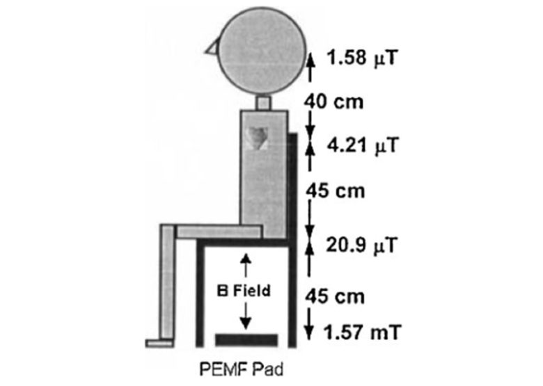
Male study participants aged 30-65 years in Australia were exposed to 50 Hz ELF-EMFs pulse-modulated at 3 Hz for 20 minutes in a sitting position. After a 20-minute break, they were again exposed for 20 minutes.
As a result, heart rate variability was altered, which showed a relative sympathetic predominance.
This was more pronounced as the time they were exposed to the EMFs increased.
The EMF generator was placed under the chair, and its strength was 1.57 μT at the head, 4.21 μT at the back, and 20.9 μT at the buttocks.
Increase in LF/HF Ratio
The LF/HF ratio increased as the duration of EMF exposure increased, indicating the relative sympathetic dominance.
Since this study is a pilot study with only five experimental participants, no statistical data analysis was performed. Data are averages of the five participants, own-calculated from the data described in the paper.
Consequences of Autonomic Imbalance
Heart rate variability was altered by EMF exposure.
They showed autonomic imbalance, mainly increased sympathetic activity, decreased parasympathetic activity, and relative sympathetic dominance.
And autonomic imbalance is known to correlate with various diseases.
Sudden Cardiac Death
Cardiac parasympathetic function is depressed in patients prone to development of sudden cardiac death. (Singer et al. 1988)
In ventricular fibrillation in the setting of cardiac ischemia, sympathetic activation is pro-arrhythmic, whereas parasympathetic activation is anti-arrhythmic. (Shen and Zipes 2014)
Ventricular fibrillation is a fatal arrhythmia and one of the most common causes of sudden cardiac death.
Sudden Infant Death Syndrome
It was shown that HF was significantly lower and LF/HF ratio higher in future sudden infant death syndrome victims than in control infants. (Franco et al. 1999)
In other words, increased sympathetic activity and decreased parasympathetic activity is a risk factor for sudden infant death syndrome.
Diabetes
Increased sympathetic activity and decreased parasympathetic activity inhibit insulin secretion in the pancreas, stimulate glucose release in the liver, and stimulate adrenal hormone secretion.
All of these factors can lead to raised blood sugar levels, which can cause diabetes.
Decreased parasympathetic activity has been observed in studies of diabetics and a population-based study also confirmed that diabetics have significantly lower parasympathetic activity than non-diabetics. (Liao et al. 1995)
Depression
Autonomic imbalance is also known to be involved in depression.
It was shown that patients with major depressive disorder showed an overall sympathetic dominance as compared with controls, with decreased parasympathetic activity. (Koschke et al. 2009)
It was also shown that a history of anxiety and depressive disorders is associated with decreased parasympathetic activity. (Jarrett et al. 2003)
Autonomic Imbalance and Various Diseases
Therefore, autonomic Imbalance due to EMF exposure, i.e., increased sympathetic activity and decreased parasympathetic activity, is likely to lead to various diseases.
Impaired Liver and Kidney Function
Next, I will present studies showing that the results of blood tests for liver and kidney function worsened in men and boys with exposure to EMF from workplaces and cell towers.

I will also present studies showing that exposure to EMFs from cell phones and other sources worsened the results of blood tests for liver and kidney function in mice.

These results suggest impaired liver and kidney function.
Blood Tests for Liver and Kidney Function
Blood tests assessing liver function include ALT, AST, ALP, GGT, bilirubin, albumin, cholesterol, and triglycerides.
Blood Tests Assessing Liver
ALT. ALT is an enzyme found in the liver that helps convert proteins into energy for the liver cells. When the liver is damaged, ALT is released into the bloodstream and levels increase. (Mayo Clinic)
AST. AST is an enzyme that helps the body break down amino acids. Like ALT, AST is usually present in blood at low levels. An increase in AST levels may mean liver damage, liver disease or muscle damage. (Mayo Clinic)
ALP. ALP is an enzyme found in the liver and bone and is important for breaking down proteins. Higher-than-usual levels of ALP may mean liver damage or disease, such as a blocked bile duct, or certain bone diseases, as this enzyme is also present in bones. (Mayo Clinic)
GGT. GGT is an enzyme in the blood. Higher-than-usual levels may mean liver or bile duct damage. This test is nonspecific and may be elevated in conditions other than liver disease. (Mayo Clinic)
Albumin and total protein. Albumin is one of several proteins made in the liver. Your body needs these proteins to fight infections and to perform other functions. Lower-than-usual levels of albumin and total protein may mean liver damage or disease. These low levels also can be seen in other gastrointestinal and kidney-related conditions. (Mayo Clinic)
Bilirubin. Bilirubin is a substance produced during the breakdown of red blood cells. Bilirubin passes through the liver and is excreted in stool. Higher levels of bilirubin might mean liver damage or disease. At times, conditions such as a blockage of the liver ducts or certain types of anemia also can lead to elevated bilirubin. (Mayo Clinic)
Cholesterol. An important function of the liver is to produce and clear cholesterol in the body. Most of the attention focused on cholesterol describes its potential for harmful health effects. But cholesterol is necessary for the creation of hormones, vitamin D, and enzymes needed for digestion. (Healthline)
In addition to liver disease, underactive thyroid, pernicious anemia, sepsis, malabsorption, etc. can cause decreased cholesterol. (Healthline)
Triglyceride. Triglycerides are the scientific term for fats. In addition to liver disease, type II diabetes, underactive thyroid, kidney disease, obesity, etc. cause increased triglyceride. (SelfDeCode)
Blood tests assesing kidney function include urea nitrogen, creatinine, and uric acid.
Blood Tests Assessing Kidney
Urea nitorogen: Urea nitrogen is a waste product. It develops when your body breaks down the protein in the foods you eat. It forms in your liver and travels through your blood to your kidneys, which then filter it out of your blood. It leaves your body through your urine. (Cleveland Cinic)
If you have too much urea nitrogen in your blood, your kidneys aren’t filtering it properly. You may have a condition that’s affecting your kidneys’ health. (Cleveland Cinic)
Creatinine : Creatinine is a chemical compound left over from energy-producing processes in your muscles. Healthy kidneys filter creatinine out of the blood. Creatinine exits your body as a waste product in urine. An increased level of creatinine may be a sign of poor kidney function. (Mayo Clinic)
Uric acid: Uric acid is a chemical produced when your body breaks down foods that contain organic compounds called purines. Most uric acid is dissolved in the blood, filtered through the kidneys, and expelled in the urine. (Healthline)
In addition to kidney disorders such as acute kidney failure, kidney stones, cancer, diabetes, gout, sepsis, malabsorption, etc. can cause increaesd uric acid. (Healthline)
Studies
Liu et al. 2013
For male workers aged 20-40 years working at a stamping workshop and a welding workshop in the automotive industry, the results of blood tests for liver function worsened when they worked in the welding workshop.
These results suggest impaired liver function.
The strength of ELF-EMFs was 0.07 μT in the stamping workshop and 0.51 μT in the welding workshop.
Blood Tests for Liver Function
Also, increases in abnormal electrocardiogram (ECG) and loss of hair were observed.
Increase in ECG abnormalities
The percentage of abnormal ECG increased 4-fold with the work at the welding shop with strong EMFs.
Increase in Loss of Hair
The reports of loss of hair increased 4-fold with the work at the welding shop with strong EMFs.
Aziz et al. 2017
For boys aged 6-12 years living in Khan Younis, Gaza Strip, Palestine, the results of blood tests for kidney and liver function worsened when they lived near a cell tower for prolonged periods.
These results suggest impaired kideny and liver function.
Also, treatment with olive oil, which has antioxidant properties, was protective in the kidneys, but both protective and counterproductive in the liver.
Blood Tests for Kidney Function
Blood Tests for Liver Function
Peighambarzadeh and Tavana 2017
Cell phones were kept in talk mode for 4 hours per day, and mice aged 1 month, equivalent to children, were exposed to their EMFs for 20 or 40 days.
As a result, the results of blood tests for liver and kidney function worsened as the exposure days increased.
These results suggest impaired liver and kidney function.
Blood Tests for Liver Function
Blood Tests for Kidney Function
Shoorei et al. 2018
Mice aged 3-4 weeks, equivalent to children, were exposed to 50 Hz ELF-EMF with a strength of 3 mT for 4 hours per day for 2 months.
As a result, reactive oxygen species (ROS) increased in the liver, and the results of blood tests for liver function worsened.
These results suggest impaired liver function.
Also, treatment with vitamin E, which has antioxidant properties, suppressed the damage above.
Increase in ROS
Increase in lipid peroxidation and decreases in total antioxidant capacity and antioxidant activity mean an increase in ROS.
Blood Tests for Liver Function
Damege to the Liver and Kidneys
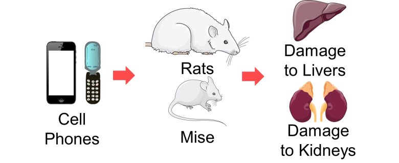
Next, I will present studies showing that exposure to EMF from cell phones and other sources damaged the livers and kidneys of rats and mice.
Studies
TOPAL et al. 2015
Pregnant rats were exposed to 900 MHz RF-EMFs with a strength of 54 μW/cm2 for 1 hour per day for 1 week in late pregnancy.
As a result, reactive oxygen species (ROS) increased in the livers of the born-son rats, damaged the livers, and hepatocytes, main cells of livers, underwent necrosis.
Increase in ROS
An increase in lipid peroxidation and a decrease in antioxidant activity mean an increase in ROS.
Damage to the Liver
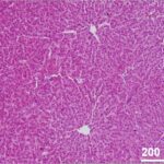
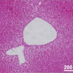
Due to the EMF exposure during pregnancy, hydropic degeneration occurred in the livers of the son rats.
Cell swelling is also known as hydropic degeneration because it is the influx of water along with sodium ions. If not stopped, acute cell swelling will cause lysis (breaking down of membrane) and death of the cell. (Miller and Zachary 2017)
Damage to the Liver (magnified)
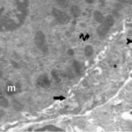
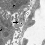
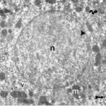
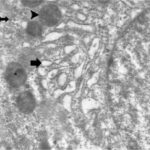
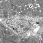
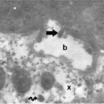
Due to the EMF exposure during pregnancy, liver cells of the son rats were damaged, and hepatocytes fragmented and underwent necrosis.
Bedir et al. 2018
Rats aged 16-20 weeks, equivalent to adolescents, were exposed to 2100 MHz RF-EMFs at a whole-body average SAR of 0.024 W/kg for 6-12 hours per day for 30 days
As a result, the results of blood tests for kidney function worsened, reactive oxygen species (ROS) in the kidneys increased, cell death in the kidneys increased, the kidneys were damaged, and renal tubules degenerated and underwent necrosis.
Renal tubules are tissues that reabsorb water and needed substances from raw urine.
Blood Tests for Kidney Function
Increase in ROS
An increase in lipid peroxidation and a decrease in antioxidant activity mean an increase in ROS.
Increase in Cell Death 1
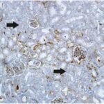
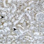
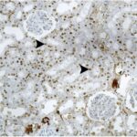
Due to the EMF exposure, cell death increased in the kidneys.
Increase in Cell Death 2
The cell death score in the kidneys increased as the duration of EMF exposure increased.
Damage to the Kidneys
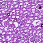
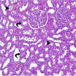
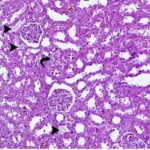

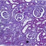
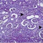
Due to the EMF exposure, renal tubules were damaged and the interstitial tissue became fibrotic (scarred).
The ductruction of normal kidney parenchyma (functional parts) due to fibrosis is a common feature in patients with chronic kidney disease. (Eddy 2014)
Degeneration of Renal Tubules
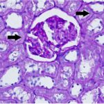
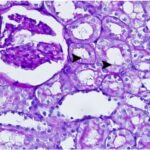
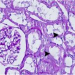
Due to the EMF exposure, renal tubules degenerated.
Necrosis of Renal Tubules
The tubular necrosis score increased as the duration of EMF exposure increased.
Acute tubular necrosis is a disease involving cell damage and death in renal tubules, but true cellular necrosis is usually minimal. (Hanif et al. 2023)
Hasan et al. 2021
Smartphones were placed on the ceilings of breeding cages and kept on incoming calls for 40 or 60 minutes per day, and male mice aged 6 weeks, equivalent to children and adolescents, were exposed to their EMFs for 60 days.
As a result, the results of blood tests for kidney function worsened, the kidneys were damaged, and inflammatory response occurred.
Blood Tests for Kidney Function
The creatinine increased 3-fold due to 60 min per day of smartphone EMF exposure; high creatinine levels suggest impaired kidney function.
Damage to the Kidneys
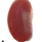
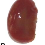
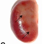
The kidneys were discolored due to 60 min per day of smartphone EMF exposure.
Damage to the Kidneys and
Inflammatory Response
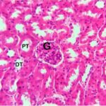
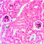
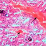
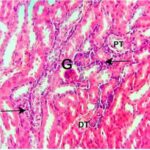
Due to the smartphone EMF exposure, the blood vessels became congested and infiltrated with immune cells, meaning inflammatory response in the kidneys.
Mortazavi et al. 2016
Cell phones with a local SAR of 1.15 W/kg were placed and kept in talk mode for 3 or 6 hours per day, and adult female rats were exposed to their EMFs at a distance of 5 cm for 7 days.
As a result, inflammatory response occurred in the liver.
Inflammatory Response in Liver
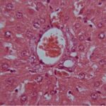
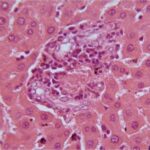
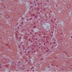
Immune cell infiltration became more pronounced as the duration of cell phone EMF exposure increased, meaning inflammatory response in the liver.
Oktem et al. 2005
Rats aged 8 weeks, equivalent to adolescents, were exposed to 900 MHz RF-EMFs for 1 hour per day for 10 days.
As a result, reactive oxygen species (ROS) increased in the kidneys, and renal tubules were damaged.
Treatment with melatonin, which has antioxidant properties, suppressed the damage above.
Increase in ROS
An increase in lipid peroxidation and a decrease in antioxidant activity mean an increase in ROS.
Damage to Renal Tubules
Urine NAG, a marker for renal tubular damage, increased 5-fold due to the EMF exposure. Melatonin, which has antioxidant properties, suppressed it.
Acute tubular necrosis is a disease involving cell damage and death in renal tubules, but true cellular necrosis is usually minimal. (Hanif et al. 2023)
Catalact and Damage to the Eyes
Next, I will present a study showing that lens opacity and retinal damage occured with exposure to EMFs from workplaces.

I will also present a study showing that cataracts increased in calves with exposure to EMFs from a cell tower.

I will also present a study suggesting that exposure to EMFs from cell phones increased reactive oxygen species (ROS) in the lenses of rats.

Mechanism of Cataracts by EMFs
A cataract is a disease where the lens of the eye becomes opaque, making objects appear blurred or hazy.
A state where ROS increased to a level that damages cells is called oxidative stress.
Opacity of the lens in cataracts is a direct result of oxidative stress. (Vinson 2006)

And numerous studies have shown that EMFs increase ROS.
Therefore, an increase in ROS due to EMF exposure is likely to cause opacity in the lens, leading to cataracts.
Studies
AURELL and TENGROTH 1973
For employees aged 40 years or younger at a factory tesing radar and other microwave euipment in Sweden, lens opacity and retinal damage increased when they were exposed to microwaves for a certain period.
Increase in Lens Opacity
The percentage of lens opacity increased 3-fold with the microwave exposure at the factory.
Increase in Retinal Damage
The percentage of retinal damage increased 7-fold with the microwave exposure at the factory.
Hässig et al. 2012
In a dairy farm in Switzerland, a large number of calves born with nuclear cataracts was reported after a cell tower had been erected in the vicinity of the farm.
Researchers found a higher risk of nuclear cataracts in the calves born there than the Swiss average.
Increase in Cataract
The odds of cataracts increased 4-fold in the dairy farm near the cell tower.
Balci et al. 2007
Cell phones with a local SAR of 1.2 W/kg were placed on top of the breeding cages and kept on incoming calls for 40 minutes per day, and female rats aged 6 weeks, equivalent to children, were exposed to their EMFs for 4 weeks.
As a result, In the corneas, ROS increased. In the lenses, both an increase and a decrease in ROS were suggested.
Treatment with vitamin C, which has antioxidant properties, suppressed the damage in the corneas.
Increase in ROS in the Corneas
Increase in ROS in the Lenses
An increase in lipid peroxidation and a decrease in antioxidant activity mean an increase in ROS.

