Cancer

It has been shown that EMF exposure increases various types of cancer, including leukemia (especially lymphoid leukemia), lymphoma, brain tumors, breast cancer, testicular cancer, lung cancer, and pancreatic cancer.
In recent years, these cancers are on the rise, and EMFs are likely to be one of the factors in their increase.
Recent Trends (*)
All Cancers :
Men
Women
Myeloid Leukemia :
Men
Women
Lymphoid Leukemia :
Men
Women
Lymphoma :
Men
Women
Breast Cancer :
Women
Testicular Cancer :
Men
Lung Cancer :
Men
Women
Pancreatic Cancer :
Men
Women
Prostate Cancer :
Men
Produced based on the data from the Cancer Incidence in Five Continents (World Health Organization), age-adjusted using the world standard population.
Table of ContentsAll_Pages
Leukemia and Lymphoma
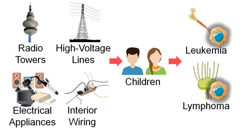
First, I will present studies showing that leukemia, particularly lymphoid leukemia, and lymphoma increased in children with exposure to EMFs from radio towers, high-voltage lines, electrical appliances, and interior wiring.
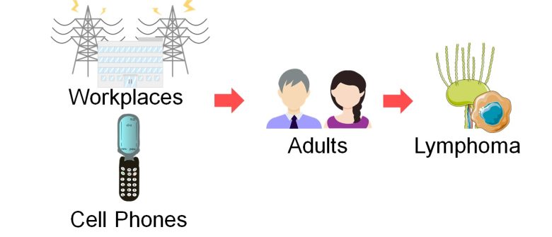
I will also present studies showing that lymphoma increased in adults with exposure to EMFs from workplaces and cell phones.
Lymphoid Leukemia and Lymphoma
Classification
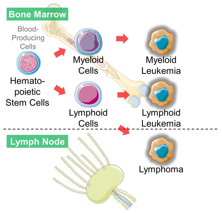
Both leukemia and lymphoma are blood cancers.
Leukemia develops in the bone marrow and is characterized by large numbers of abnormal blood cells in the peripheral blood.
Lymphoma develops in the lymph nodes and is often accompanied by swollen lymph nodes with no abnormal blood cells in the peripheral blood.
Leukemia is largely divided into myeloid leukemia and lymphoid leukemia according to the type of blood cells that have become cancerous.
Lymphoid leukemia and lymphoma are the same in that lymphocytes (a type of white blood cells) or their precursors have become cancerous, and the boundary between the two is ambiguous.
For example, acute lymphoblastic leukemia is also known as lymphoblastic lymphoma, which is a leukemia if a large percentage of tumor cells arise in the bone marrow, and a lymphoma otherwise. According to the WHO classification, they are the same disease. (Alaggio et al. 2022)
Recent Trends
In developed countries, while the incidence of myeloid leukemia has not changed significantly, both lymphoid leukemia and lymphoma are on the rise (*).
Produced based on the data from the Cancer Incidence in Five Continents (World Health Organization), age-adjusted using the world standard population.
Trends in Myeloid Leukemia
Trends in Lymphoid Leukemia
Trends in Lymphoma
Involvement of EMFs
And it has been shown that EMF exposure increases especially lymphoid leukemia among leukemias, and lymphoma.
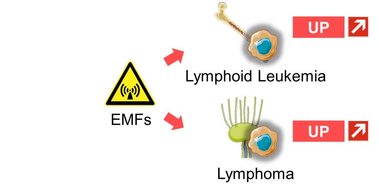
That is, EMFs may be creating the upward trends in lymphoid leukemia and lymphoma.
Studies
Michelozzi 2002
The Vatican Radio station, located at the northwest edge of Rome, Italy, is a very powerful station that transmits all over the world.
The concern of the population regarding possible health effects associated with the station had been raised.
The regional government requested epidemiologic investigations, and this study was undertaken.
The study found that for children aged under 15 years, leukemia increased as the distance from their homes to the radio station decreased.
An increase in deaths from leukemia in adults was also observed.
Increase in Leukemia
Dolk et al. 1997
At that time, the Guardian newspaper reported a claim from a doctor of Birmingham about excessed cases of leukemia and lymphoma near the Sutton Coldfield radio and television transmitter in the West Midlands, England.
Following the claim, the Small Area Health Statistics Unit in the United Kingdom was asked to investigate the situation, and this study was undertaken.
The study found that leukemia, especially lymphoid leukemia, increased as the distance from their homes to the radio and TV transmitter decreased in residents aged 15 years and older.
Increase in Lymphoid Leukemia
SAVITZ et al. 1988
For children aged under 14 years in the Denver metropolitan area, childhood cancer increased as the strength of EMFs from interior wiring increased, particularly acute lymphocytic leukemia and lymphoma.
Increase in Childhood Cancer
The odds of childhood cancer increased 2-fold with an average of 0.2 μT of the EMFs from interior wiring.
The weakest group of interior wiring is excluded since its EMF strength is almost the same as that of buried wiring.
Acute Lymphocytic Leukemia
and Lymphoma
The odds of acute lymphocytic leukemia and lymphoma in children increased 3-fold with an average of 0.2 μT of the EMFs from interior wiring.
Olsen et al. 1993
For children aged under 15 years throughout Denmark, childhood cancer increased when the strength of ELF-EMFs from high-voltage lines and high-voltage substations was stronger.
Increase in Childhood Cancer
The odds of leukemia and brain tumors in children increased 6-fold and the odds of lymphoma in children increased 5-fold with 0.4 μT or more of the ELF-EMFs from high-voltage lines and high-voltage substations.
Schroeder and Savitz 1997
For male employees of five large U.S. power companies, lymphoma increased when cumulative exposure to ELF-EMFs at the workplaces was higher.
This was especially more pronounced when looking at the cumulative exposure over the past 10-20 years.
Increase in Lymphoma
Also, among lymphoma, high-grade lymphoma increased even more.
This was also more pronounced when looking at the cumulative exposure over the past 10-20 years.
Increase in
High-Grade Lymphoma
Also, lymphoma increased even more among employees whose hiring year was five years late.
Increase in Lymphoma
Among Younger Employees
The mortality rate of lymphoma increased 5-fold in the middle 70-89% for cumulative exposure to the ELF-EMFs at the workplaces.
Feychting and Alhbom 1993
For children aged 16 years or younger living within 330 yards (300 m) of high-voltage lines throughout Sweden, leukemia increased as the distance from their homes to the high-voltage lines decreased and as the strength of their ELF-EMFs increased.
Increase in Leukemia
Schüz et al. 2001
For children aged 15 years or younger throughout the former West Germany, acute leukemia increased as the strength of ELF-EMFs in bedrooms at night increased.
Increase in Acute Leukemia
The odds of acute leukemia in children increased 5-fold with 0.4 μT or more of the ELF-EMFs in the bedrooms at night.
Also, acute leukemia increased even more among infants and toddlers aged 4 years or younger.
Increase in Acute Leukemia
Among Infants & Toddlers
The odds of acute leukemia in infants and toddlers increased 15-fold with 0.4 μT or more of the ELF-EMFs in the bedrooms at night.
Also, for children aged 15 years or younger, acute leukemia increased even more when the children resided in the same house from birth, i.e., when they had been exposed to ELF-EMFs from infancy and toddlerhood.
Increase in Acute Leukemia
Resided in the Same House
The odds of acute leukemia in children increased 16-fold with 0.4 μT or more of the ELF-EMFs in the bedrooms at night.
Kabuto et al. 2006
For children aged 15 years or younger in Tokyo, Nagoya, Kyoto, Osaka and Kitakyushu metropolitan areas in Japan, acute leukemia, especially acute lymphoblastic leukemia, increased when the strength of ELF-EMFs in bedrooms at night were stronger.
Increase in
Acute Lymphoblastic Leukemia
Dr. Kabuto's study, which began at the request of the WHO, was highly praised by the WHO, but was ignored by the Japanese government as unreliable. The government's evaluation committee gave it a "lowest grade" of C (not good) for all 12 categories on a scale of three ABCs. (Ogino 2019)
Dr. Kabuto lamented, "Why am I being treated like this when my results were similar to those of Western studies?" He died of lymphoma shortly after submitting his report. (Ogino 2019)
Oddly enough, the cause of death was the same lymhpoid cancer as acute lymphoblastic leukemia, which Dr. Kabuto discovered a strong correlation with EMFs in his study.
There seems to be political pressure in Japan toward studies on the biological effects of EMFs.
Hatch et al. 1998
For children aged 14 years or younger in Illinois, Indiana, Iowa, Michigan, Minnesota, New Jersey, Ohio, Pennsylvania, and Wisconsin, acute lymphoblastic leukemia increased as the years of use of electrical appliances (EAs) increased.
Increase in
Acute Lymphoblastic Leukemia
Using EAs
Also, acute lymphoblastic leukemia increased as the TV viewing time per day increased and as the TV viewing distance decreased.
Increase in
Acute Lymphoblastic Leukemia
Using TVs
The odds of acute lymphoblastic leukemia increased 4-fold with the TV viewing time of more than 6 hours per day and with the TV viewing distance of less than 1.3 yards (1.2 m).
Also, for pregnant women, acute lymphoblastic leukemia in children born to them increased when they had used electrical appliances.
Increase in
Acute Lymphoblastic Leukemia
Using EAs During Pregnancy
Also, acute lymphoblastic leukemia in children born to them increased as the TV viewing distance during pregnancy decreased.
Increase in
Acute Lymphoblastic Leukemia
Using TVs During Pregnancy
The odds of acute lymphoblastic leukemia in children increased 2-fold with the TV viewing distance of less than 1.3 yards (1.2 m).
Moscow Signal
Here is presented an episode from the Cold War era called Moscow Signal, from Reviews on Environmental Health, a quarterly peer-reviewed review journal covering the field of environmental health.
Martínez 2019
The 10-story US embassy in Moscow was irradiated with microwaves (*) by the Soviet government from 1953 to April, 1979.
RF-EMFs in the frequency range used by radios, cell phones, Wi-Fi, etc.
It is estimated that the strength of the microwaves was about 5 μW/cm2 and irradiated for 9 hours per day.
This radiation attack soon became known as the “Moscow Signal.” However, the US government decided to keep it a secret until 1972, when they began to inform some of the embassy workers. The other members of staff at the building were not informed of the facts until 1976.
Indeed, it was not until the beginning of 1976 when the event came to light, in an article published in Time magazine, which reported that many members of the embassy staff had returned to the US with severe health problems, that two ambassadors had died of cancer, and that a third was suffering from leukemia.
On June 21, 1976, Dr. Lilienfeld and his team signed a contract with the U.S. government for an epidemiological study that took two years and resulted in a 400-page report.
The medical records showed a significant increase in appendicitis, sleepwalking, venereal disease, protozoal intestinal disease, benign neoplasms, diseases of the nerves and peripheral ganglia and complications during pregnancy, childbirth and puerperium.
The health questionnaires showed a significant increase in depression, irritability, difficulty in concentration, memory loss, eye problems, psoriasis, skin conditions, anemia, ulcers, and other symptoms, many of which were related to electromagnetic hypersensitivity.
In addition, there was an increase in the number of deaths from leukemia and breast cancer.
Increase in Leukemia and Breast Cancer
At the Moscow embassy, the number of observed deaths of leukemia was 3 times higher than the national average, and for breast cancer 5 times.
Although this was not statistically significant, it did not include the third ambassador's death from leukemia, and is suspected to have suppressed the number of deaths from breast cancer.
Also, Dr. Lilienfeld indicated that because of the sample size limitations, the study was not able to significantly detect increased risks unless they were unusually large.
An initial study of 43 people conducted in 1967 found that 20 out of 37 people in the exposed group had abnormal chromosomes, and 2 out of 7 people in the unexposed group had abnormal chromosomes. (1.8 times more chromosome abnormalities)
Another study conducted in 1976 found higher white blood cell count among the Moscow embassy employees compared to other employees of the foreign affairs service.
It suggests that the biological effects of EMFs had already been discovered at least as early as the 1950s and 1960s.
The danger of the disease-provoking capacity (e.g. cancer) of EMFs being used as anti-personnel weapons is apparent.
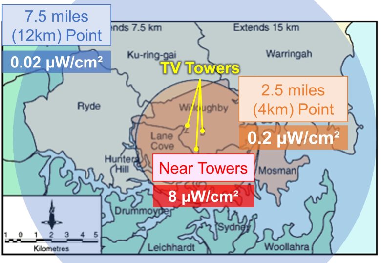
The strength of the irradiated microwaves is 5 μW/cm2, which is not much different from the strength of EMFs exposed in the vicinity of radio towers and cell towers (data are shown on page 5).
Abnormal Blood Counts
Next, I will present studies showing that exposure to EMFs from cell phones and other sources caused abnormalities in red blood cell count, platelet count, and white blood cell count in rats and mice.
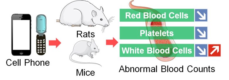
Abnormal Blood Counts by Leukemia
With leukemia, blood tests will likely show higher than usual white blood count, which includes leukemic cells, and may also show lower than usual red blood cell and platelet cell counts. (healthline)
While leukemia typically leads to a high white blood cell count, it can sometimes lead to low white blood cell counts. (myleukemiateam)
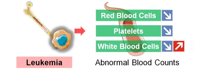
And, it has been shown that exposure to EMFs decreases blood cell count and platelet count and increases or decreases white blood cell count in rats and mice.
Studies
Hasan et al. 2021
Smartphones were placed on the ceilings of breeding cages and kept on incoming calls for 40 or 60 minutes per day, and male mice aged 6 weeks, equivalent to children/adolescents, were exposed to their EMFs for 60 days.
As a result, red blood cell (RBC) count decreased and white blood cell (WBC) count increased as exposure time per day increased.
Decrease in RBC and
Increase in WBC
Alghamdi and El-Ghazaly 2012
Cell phones with a local SAR of 0.49 W/kg were placed inside breeding cages, and male mice aged 6 weeks, equivalent to children/adolescents, were exposed to their EMFs for 15-60 minutes per day for 2 weeks.
As a result, red blood cell (RBC) count and platelet (PLT) count decreased and white blood cell (WBC) count increased as the exposure time per day increased.
Also, various abnormalities in red blood cells were observed in blood smears.
Decreases in RBC, PLT and
Increase in WBC
The white blood cell count value is unnatural, but it is not clear if it is a typo or if there is some other reason.
Abnormalities in Red Blood Cells

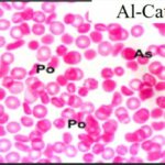
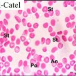

Due to the exposure to the cell phone EMFs, various abnormalities occurred in red blood cells. It can also be seen that the longer the exposure time, the lower red blood cell count.
El-Bediwi et al. 2012
Test cell phones with a local SAR of 0.96 W/kg were placed in the center of breeding cages, and male rats aged 3 months, equivalent to adolescents, were exposed to their EMFs for 1 hour per day for 3 or 6 months.
As a result, red blood cell (RBC) count, platelet (PLT) count, and white blood cell (WBC) count decreased as the exposure duration increased.
Decreases in RBC, PLT, WBC
Jelodar et al. 2010
Male pup rats were exposed to RF-EMFs similar to those emitted from cell towers for 5 hours a day for 70 days.
As a result, red blood cell (RBC) count, platelet (PLT) count, and white blood cell (WBC) count decreased.
Decreases in RBC, PLT, WBC
Brain Tumors
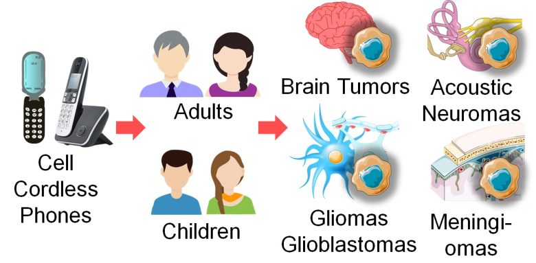
Next, I will present studies showing that brain tumors, especially gliomas, and more especially glioblastomas, the most malignant brain tumors, as well as meningiomas and acoustic neuromas, inner ear tumors, increased in adults and children with exposure to EMFs from cell/cordless phones.
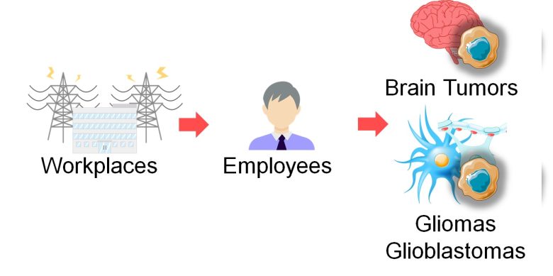
I will also present studies showing that brain tumors, gliomas, and glioblastomas increased with exposure to EMFs from workplaces.
EMF Absorption and Brain Tumors
Children Absorb EMFs Throughout Their Heads
Since children's heads are smaller than adults', they have been shown to absorb more EMF energy. (Gandhi et al. 1996)
The following images compare the distributions of specific absorption rate (SAR), representing the amount of absorbed EMF energy, between children and adults when an 835 MHz, 0.6 W EMF emitter that imitates cell phones is held against the ear tilted at 30°. (Gandhi et al. 1996)
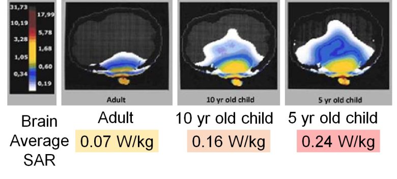
You can see that children absorbed EMFs throughout their heads unlike adults, and that the brain average EMF absorption was several times higher than that of adults.
In Children, Tumors Occur on Both Sides of the Brain
The adult use of cell phones has been shown to increase brain tumors, primarily on the use side of cell phones.
On the other hand, the child use of cell phones has been shown to increase brain tumors not only on the use side but also on the opposite side.
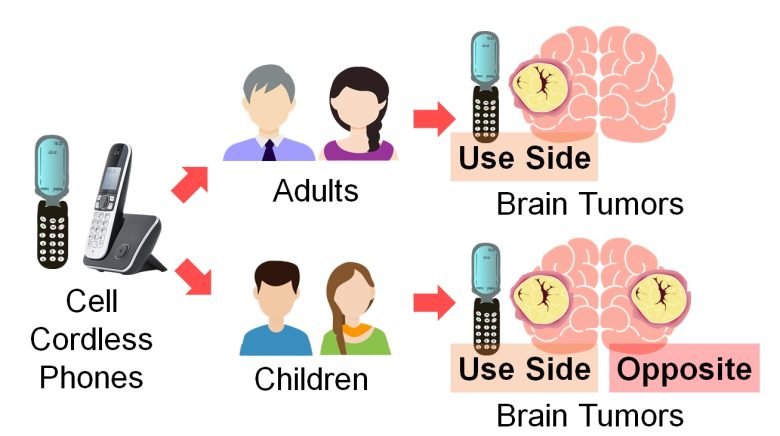
This is likely largely due to the difference in EMF absorption in the brain between adults and children.
Cell Phones and Glioblastomas
What is Glioblastomas?
In addition to neurons, which are responsible for information processing in the brain, there are cells that support the activity of neurons, called glial cells, such as astrocytes and oligodendrocytes.
Cancer developing from these glial cells is called gliomas.
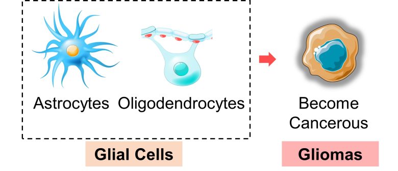
Gliomas are the most common malignant tumours of the brain, accounting for over 80% of all primary brain malignancies. (Persaud-Sharma et al. 2017)
Of these gliomas, grade IV are called glioblastomas and are known as the most malignant brain tumors, with five-year survival rates of less than 10%.
Recent Trends
In the United States and the United Kingdom, among gliomas, there is a trend toward an increase in grade IV, glioblastomas, while toward a decrease in grade III or lower.
And this trend began with the launch of cell phone commercial service.
Trend in Gliomas in the U.S.
From 1981 to 1996, the incidence rate of glioma grade IV (glioblastoma) increased by 40% in 15 years, while grade III or lower decreased by 30% conversely. (Li et al. 2018)
Trend in Gliomas in the U.K.
From 1985 to 2010, the new cases of glioma grade IV (glioblastoma) increased 6-fold in 25 years, while grade III or lower decreased by 60% conversely. (de Vocht 2016)
Involvement of Cell Phones
And, it has been shown that cell phone use increases the risk of grade IV gliomas (glioblastomas) while conversely decreasing those of grades I and II.
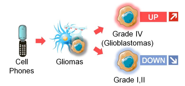
That is, cell phones may be creating an upward trend in grade IV gliomas (glioblastomas) and a downward trend in lower-grade gliomas.
Studies
Aydin et al. 2011
For children aged 19 or younger in Denmark, Sweden, Norway, and Switzerland, brain tumors increased as the years of cell phone use increased and as the call time and the number of calls on their cell phones increased.
The increase in brain tumors was observed on both the use side of cell phones and the opposite side.
Increase in Tumors
(Years of Use)
The odds of brain tumors in children increased 4-fold on both the use and opposite side of cell phones with more than 4 cumulative years of cell phone use.
Number of calls was less than once a week.
Increase in Tumors
(Call Time)
The odds of brain tumors in children increased 3-fold on the use side of cell phones and 6-fold on the opposite side with more than 144 hours of cumulative call time on the cell phones.
Number of calls was less than once a week.
Increase in Tumors
(Number of Calls)
The odds of brain tumors in children increased 3-fold on the use side of cell phones and 5-fold on the opposite side with more than 2638 of cumulative number of calls on cell phones.
Number of calls was less than once a week.
Hardell et al. 2006
For residents aged 20 years or older in central Sweden, malignant brain tumors, especially high-grade gliomas, increased as the years of cell/cordless phone use increased.
Increase in Malignant Brain Tumor
Also, the increases in malignant brain tumors and high-grade gliomas were greater on the use side of cell/cordless phones and smaller on the opposite side.
Increase in Malignant Brain Tumor
(Use Side and Opposite Side)
Hardell and Carlberg 2015
For residents aged 18 years or older throughout Sweden, gliomas increased as the years of cell/cordless phone use increased.
By cell phone generations, the third generation showed the greatest increase in gliomas.
Increase in Gliomas
The odds of gliomas increased as the years of use increased.
Increase in Gliomas by Generations
Also, gliomas increased on the use side of cell phones when they first used their cell phones as adults, but increased not only on the use side but also on the opposite side when they had already used their cell phones as minors.
Increase in Gliomas on Use Side
The odds of gliomas increased on the use side of cell phones with 1 year or more of cell phone use, regardless of the age at first use.
Increase in Gliomas on Opposite Side
The odds of gliomas increased on the opposite side of cell phones with 1 year or more of cell phone use, only when the age at first use was underage.
Carlberg and Hardell 2014
For patients with glioblastomas (grade IV gliomas) aged 18 years or older throughout Sweden, survival rates decreased as the years of cell phone use until the onset increased.
Increase in Mortality Hazard
By Years of Use
The mortality hazard of glioblastomas increased 2.1-fold with more than 15 years of cell phone use.
An increase in the mortality hazard means a decrease in the survival rate per unit of time (e.g., per mounth).
For your reference, I applied the above mortality hazard ratios to the survival rates of glioblastomas by month in the United States for 2000-2003 (Johnson and O’Neill 2011) to produce the following graph.
Decrease in Survival Rates
By Years of Use
If glioblastoma patients had been using cell phones for more than 15 years by the onset, the 5-year survival rate decreased from 5% to 0.2%. In other words, the chance of survival almost disappeared.
Also, survival rates decreased as the age at first use of cell phones decreased.
Increase in Mortality Hazard
By Age at First Use
The mortality hazard of glioblastomas increased 2.3-fold with the use of cell phones from the age of a minor.
For your reference, I applied the above mortality hazard ratios to the survival rates of glioblastomas by month in the United States for 2000-2003 (Johnson and O’Neill 2011) to produce the following graph.
Decrease in Survival Rates
By Age at First Use
If glioblastoma patients had been using cell phones since they were minors, the 5-year survival rate decreased from 5% to 0.1%. In other words, the chance of survival almost disappeared.
Glioblastomas are grade IV gliomas, but patients with grade I-II gliomas conversely increased survival rates with cell phone use.
Decrease in Mortality Hazard
for Grade I-II
The mortality hazard of grade I-II gliomas decreased by 40% with 1 year or more of cell phone use.
For your reference, I applied the above mortality hazard ratios to the survival rates of grade I-II gliomas by month in the United States for 2000-2011 (Claus et al. 2015) to produce the following graphs.
Increase in Survival Rates
for Grade I-II
If grade I-II glioma patients had been using cell phones for 1 year or more by the onset, the 5-year survival rate increased conversely from 62% to 75%.
Carlberg et al. 2017
For residents aged 18 years and older throughout Sweden, glioblastomas (grade IV gliomas) increased as cumulative exposure to ELF-EMFs at the workplaces increased.
This was more pronounced when looking at the cumulative exposure over the past 1-14 years.
Increase in Glioblastoma
On the other hand, grade I-II gliomas conversely decreased.
Decrease in Grade I-II Gliomas
The odds of grade I-II gliomas decreased conversely with the EMF exposure at the workplaces.
HARDELL et al. 2013
For residents aged 18 years and older throughout Sweden, acoustic neuromas, inner ear tumors, increased as the years of cell/cordless phone use increased.
Increase in Acoustic Neuromas
By cell phone generations, the third generation showed the greatest increase in acoustic neuromas.
Increase in Acoustic Neuromas
by Generations
The odds of acoustic neuromas increased 4-fold with 1-5 years of third-generation cell phone use.
Coureau et al. 2014
For residents aged 16 years or older in 4 departments (Gironde, Calvados, Manche, Hérault) in France, gliomas and meningiomas increased when they used cell phone.
Increase in Gliomas
The odds of gliomas increased 2-fold with 10 years or more of cell phone use, 4-fold with more than 15 hours of average monthly call time, and 3-fold with 896 hours or more of cumulative call time.
Number of calls was less than once a week.
Increase in Meningiomas
The odds of meningiomas increased 2-fold with 10 years or more of cell phone use, 2-fold with more than 15 hours of average monthly call time, and 3-fold with 896 hours or more of cumulative call time.
Number of calls was less than once a week.
Users with 896 hours or more of cumulative call time were defined as heavy users, and their cell phone usage and tumor locations were examined in detail.
The heavy users showed increases in gliomas and meningiomas in the temporal lobe, which is in close proximity to the cell phone during use.
Heavy Users and Tumor Locations
For the heavy users, the odds of gliomas increased 4-fold and meningiomas 8-fold in the temporal lobe.
Number of calls was less than once a week.
Also, the heavy users showed a considerable increase in gliomas when using cell phones only in urban areas.
Heavy Users in Urban Areas
For the heavy users who used their cell phones only in urban areas, glioma increased 8-fold and meningioma 3-fold.
Number of calls was less than once a week.
Breast Cancer
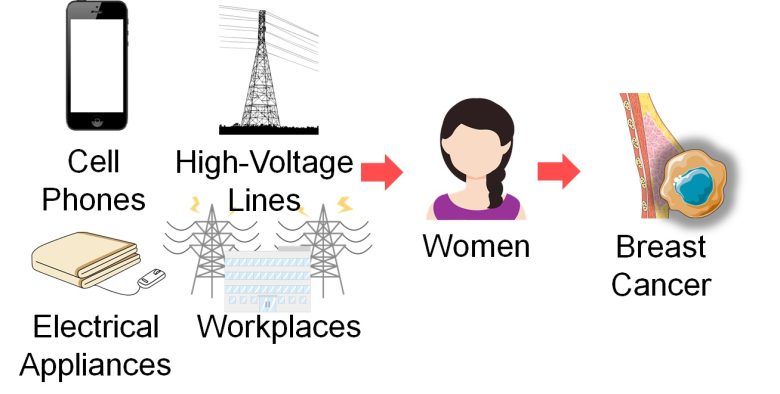
Next, I will present studies showing that breast cancer increased in women with exposure to EMFs from cell phones, high-voltage lines, electrical appliances, and workplaces.
ER-Positive/Negative Breast Cancer
Classification
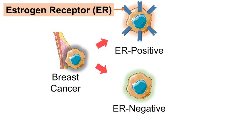
Breast cancer can be divided into two categories: cancers that receive and grow on female hormones and those that do not. These hormones are estrogen and progesterone.
Those with receptors for estrogen (ER) are called ER-positive breast cancer and those without are called ER-negative breast cancer.
ER-positive breast cancer accounts for 70% of all breast cancers. (Gombos 2019)
Recent Trends
In the United States, among women aged under 50 years, ER-positive breast cancer is on the rise while ER-negative breast cancer is on the decline.
On the other hand, among women aged 50 years or older, there has been no significant change in ER-positive breast cancer while ER-negative breast cancer is on the decline, but the decrease is smaller than among younger women.
Trends in Breast Cancer
Aged Under 50 Years
In the 16 years from 1992 to 2008, among women aged 30-49 years, the incidence of ER-positive breast cancer increased by 20% while the incidence of ER-negative breast cancer decreased by 30%. (Anderson et al. 2011)
Trends in Breast Cancer
Aged 50 Years or Older
In the 16 years from 1992 to 2008, among women aged 50-84, there was no significant change in the incidence of ER-positive breast cancer while the incidence of ER-negative breast cancer decreased by only 10%. (Anderson et al. 2011)
Involvement of EMFs
And it has been shown that EMF exposure tends to increase ER-positive breast cancer in women aged roughly under 50 years, and conversely ER-negative breast cancer in women aged roughly over 50 years.
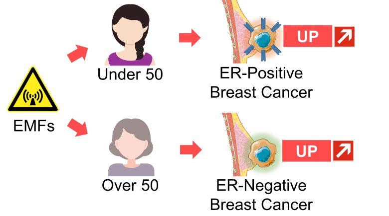
That is, EMF may be creating an upward trend in ER-positive breast cancer in women aged under 50 years and a smaller downward trend in ER-negative breast cancer in women aged 50 years or older.
Studies
Shih et al. 2020
For women aged 20 years or older recruited at the breast surgery outpatient department of a medical university hospital, Taiwan, breast cancer increased when the time spent on their smartphones before sleep was longer.
This was especially more pronouced among women with smartphone addiction.
Increase in Breast Cancer
Using Before Sleep
The odds of breast cancer increased 5-fold with more than 4.5 minutes of smartphone use time before sleep, and even 7-fold among the women with smartphone addiction.
Also, breast cancer increased when they used their smartphones near the chest.
Increase in Breast Cancer
Using Near the Chest
The odds of breast cancer increased 2-fold with hand-held use of smartphones, i.e., use near the chest.
Also, breast cancer increased when they carried their smartphones near the chest.
Increase in Breast Cancer
Carrying Near the Chest
The odds of breast cancer increased 5-fold with a smartphone carried near the chest.
Zhu 2003
For African-American women aged 20-64 years in three Tennessee counties (Davidson, Shelby, and Hamilton counties), breast cancer increased as the years of use of electric bedding (electric blankets, electric mattress pads, and heated waterbeds) increased.
This was especially more pronounced among premenopausal, younger women.
Increase in Breast Cancer
The odds of breast cancer increased 5-fold with more than 10 years of electric bedding use, and 8-fold among premenopausal women.
Feychting et al. 1998
For women aged 16 years or older living within 330 yards (300 m) of high-voltage lines throughout Sweden, breast cancer increased as the distance from their homes to the high-voltage lines increased and as the strength of ELF-EMFs from the high-voltage lines increased and as their cumulative exposure increased.
The above trends were observed primarily among younger women aged under 49 years.
Increase in Breast Cancer
Among breast cancers, there was a further increase in ER-positive breast cancer, which was more pronounced among those aged 49 years or younger.
Increase in ER-Positive Breast Cancer
The risk of ER-positive breast cancer in women aged 49 years or younger increased 7-fold with 0.1 μT or more of the ELF-EMFs from the high-voltage lines.
Kliukiene et al. 2003
For female radio and telegraph operators throughout Norway, breast cancer increased as cumulative exposure to ELF-EMFs and RF-EMFs at workplaces increased.
Increase in Breast Cancer
The odds of breast cancer increased 2-fold in the top 33% for the cumulative exposure to EMFs at the workplaces.
Also, among women aged under 50 years, ER-positive breast cancer increased as cumulative EMF exposure increased.
Increase in ER-Positive Breast Cancer
The odds of ER-positive breast cancer increased 2-fold in the top 33% for the cumulative exposure to EMFs at the workplaces.
On the other hand, among women aged 50 years or older, ER-negative breast cancer increased as cumulative EMF exposure increased.
Increase in ER-Negative Breast Cancer
The odds of ER-negative breast cancer increased 8-fold in the top 33% for the cumulative exposure to EMFs at the workplaces.
Demers et al. 1991
For male employees in areas covering about 15% of the U.S. population, exposure probability to ELF-EMFs was classified by occupations into one of five categories: none, lower, likey, likeky including RF-EMFs, and definite.
Male breast cancer increased as the exposure probability to ELF-EMFs increased.
Increase in Male Breast Cancer
By Exposure Probability
The odds of male breast cancer increased 6-fold with "definite" exposure possibility to ELF-EMFs.
Also, male breast cancer increased as years of service in occupations exposed to ELF-EMFs (*) increased.
Exposure possibility was other than "none."
Increase in Male Breast Cancer
By Years of Service
The odds of breast cancer increased 2-fold with more than 30 years of service in the occupations exposed to EMFs.
Testicular Cancer
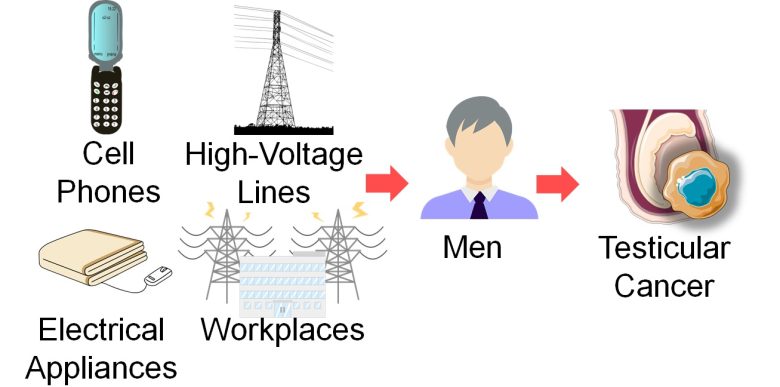
Next, I will present studies showing that testicular cancer increased in men with exposure to EMFs from cell phones, high-voltage lines, electrical appliances, and workplaces.
Seminomas and Non-Seminomas
Classification
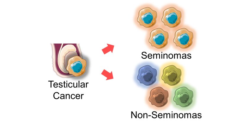
The majority of testicular cancers originate from germ cells (*), 60% of which are seminomas, 30% are non-seminomas, and the remaining 10% are mixtures of the two. (Singh et al. 2011)
e.g., spermatogonia, the stem cells of the testes
Seminomas are homogeneous tumors in which germ cells become cancerous, resembling primordial germ cells. (Singh et al. 2011)
Non-seminomas, on the other hand, are higly heterogeneous tumors that display various stages of differentiation ranging from undifferentiated cells to highly differentiated cells. (Singh et al. 2011)
Tumor heterogeneity is associated with poor prognosis and outcome and is thought that one of the leading determinants of therapeutic resistance and treatment failure. (Ramón y Cajal et al. 2020)
Recent Trends
In developed countries, both seminomas and non-seminomas are on the rise.
Trends in Seminomas
In the 25 years from 1975 to 2000, the incidence of seminoma increased by 60% (15 years) in Australia, 90% in France, and 80% in the U.S., with no change in Japan (*). (Chia et al. 2010)
Trends in Non-Seminomas
In the 25 years from 1975 to 2000, the incidence of seminoma increased by 60% (15 years) in Australia, 110% in France, and 40% in the U.S., with no change in Japan (*). (Chia et al. 2010)
In Japan, testicular cancer is on the rise that began after 2000.
Involvement of EMFs
And, it has been shown that EMF exposure tends to increase both seminomas and non-seminomas, and if anything, non-seminomas.
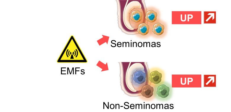
That is, EMFs may be creating the upward trends in seminomas and non-seminomas.
Studies
Stenlund and Floderus 1997
For male employees aged 20-64 years in 11 counties of the middle of Sweden, testicular cancer increased as the strength of ELF-EMFs at the workplaces increased.
This was especially more pronounced among men aged 40 years or younger.
Increase in Testicular Cancer
Also, among men aged 40 years or younger, especially non-seminomas increased.
Increase in Non-Seminomas
The odds of non-seminomas increased 16-fold with 0.4 μT or more of the ELF-EMFs at the workplaces.
Hardell et al. 2006
For men aged 20-75 years throughout Sweden, testicular cancer, especially seminomas, increased as the years of cell phone use increased.
Increase in Testicular Cancer
With Prolonged Use
Also, testicular cancer increased when they carried their cell phones near their testes.
Increase in Testicular Cancer
Carrying Near the Testes
The odds of testicular cancer increased by 30-80% with 1 year or more of cell phone use and with a cell phone carried in a pants pocket or a waist-belt pocket.
Davis and Mostofi 1993
A culuster of testicular cancer occured in two separate police departments serving geographically contiguous counties in the north-central United Stetes.
All of the affected men used radar guns (for speed enforcement) on duty, and all of them also carried the radar guns routinely with the power on near their testes, which was the only shared risk factor.
A radar gun emits EMFs onto a target and calculates the speed based on the difference in frequency between the reflected back EMFs and the original EMFs.
The study found that the number of cases of testicular cancer in the said police departments is higher than the national average.
Increase in Testicular Cancer
The number of observed cases of testicular cancer was 7 times higher than the national average in the said police departments.
Baumgardt-Elms et al. 2004
For men aged 15-69 years in Hamburg, Germany, testicular cancer increased as cumulative exposure to ELF-EMFs from high-voltage lines increased.
This was especially more pronounced among men aged 40 or younger.
Increase in Testicular Cancer
The odds of testicular cancer increased by 70% with high cumulative exposure to the high-voltage line EMFs and by 90% among men aged 40 years or younger.
VERREAULT et al. 1990
For men aged 20-69 years in 13 counties of western Washington, non-seminomas increased and seminomas decreased conversely as years of electric blanket use increased.
Increase in Non-Seminomas
The incidence rate of non-seminomas increased by 80% with more than 2 years of electric blanket use, and seminomas decreased by 20% conversely.
Other Cancers
Next, I will present studies showing that cancers increased in residents with exposure to EMFs from cell towers.

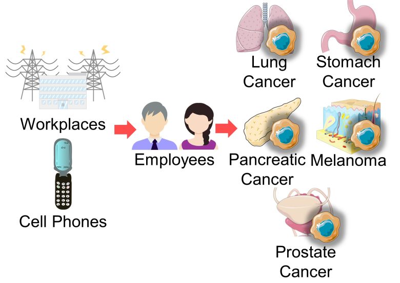
Also, I will present studies showing that various types of cancer, including lung cancer, stomach cancer, pancreatic cancer, melanoma, prostate cancer, and others, increased with exposure to EMFs from workplaces and cell phones.
Also, I will present studies showing that neuroblastoma increased in children with exposure of their parents to EMFs at workplaces.

Studies
Eger et al. 2004
For residents in Naila of Upper Franconia, Bavaria, Germany, cancers increased when they lived near a cell tower.
This was especially more pronounced with the prolonged years of residence.
Increase in Cancers
The risk of cancers increased 3-fold with 5-10 years of residence within 440 yards (400 m) of the cell tower.
Wolf and Wolf 2004
For residents in Netanya, Israel, cancers increased when they lived near a cell tower.
The average strength of EMFs within 380 yards (350 m) of the cell tower was 0.53 μW/cm2.
Increase in Cancers
The number of observed cases of cancers was four times higher than the national average within 380 yards (350 m) of the cell tower.
Tomenius 1986
For residents aged 18 years or younger in the county of Stockholm (city of Stockholm plus 22 other neighboring communities), Sweeden, childhood tumors increased when ELF-EMFs at entrance doors of their houses were stronger.
This was especially more pronounced when living in permanent dwellings, which suggests an increased risk due to prolonged EMF exposure.
Increase in Childhood Tumors
The risk of childhood cancer increased 2-fold with 0.3 μT or more of the ELF-EMFs at the entrance doors of their houses, and 4-fold especially in the permanent dwellings.
Armstrong et al. 1994
For employees of 2 power companies in France and Canada, lymphoma, lip/oral/throat cancer, stomach cancer, lung cancer, etc., increased as cumulative exposure to ELF-EMFs at workplaces increased.
Increase in Cancers
The odds of cancers increased by 40% in the top 10% for cumulative exposure to ELF-EMFs at the workplaces.
Increase in Lymphoma
The odds of lymphoma increased 2-fold in the top 10% for cumulative exposure to ELF-EMFs at the workplaces.
Increase in Lip/Oral/Throat Cancer
The odds of lip/oral/throat cancer increased 5-fold in the top 10% for cumulative exposure to ELF-EMFs at the workplaces.
Increase in Stomach Cancer
The odds of stomach cancer increased 5-fold in the middle 70-90% for cumulative exposure to ELF-EMFs at the workplaces.
Increase in Lung Cancer
The odds of lung cancer increased 10-fold in the top 10% for cumulative exposure to ELF-EMFs at the workplaces.
De Roos et al. 2001
For children aged under 19 years at 139 hospitals in the United States and English-speaking Canada, exposure probability to RF-EMFs for the parents in two years before birth was classified into three categories: none, possible, and probable.
Neuroblastoma increased in their children as the exposure probability to RF-EMFs for the parents, especially the mothers, increased
Increase in Neuroblastoma
Also, when looking at each exposure source to EMFs for the fathers at the workplaces, an increase in neuroblastoma in children was observed with exposure of fathers to EMFs from battery-powered forklifts, welding machines, and mobile radio transmitters.
Increase in Neuroblastoma
The odds of neuroblastoma in children increased 2-fold with exposure of fathers to EMFs from battery-powered forklifts, welding machines, and mobile radio transmitters.
Ji et al. 1999
For male employees aged 30-74 years in Shanghai, China, pancreatic cancer increased as the strength of ELF-EMFs at workplaces increased.
Increase in Pancreatic Cancer
The odds of pancreatic cancer increased 3-fold with "high" strength of ELF-EMFs at the workplaces.
Also, when looking at occupations exposed to EMFs, pancreatic cancer increased among electricians, and it was more pronounced as the years of service increased.
Increase in Pancreatic Cancer
Among Electrician
The odds of pancreatic cancer increased 9-fold for electricians with 35 years or more of service.
Hardell et al. 2011
For men aged 20-77 years throughout Sweden, melanoma increased as the age at first use of cell/cordless phones decreased.
This was especially more pronounced in the head and neck region, which are in close proximity to a cell/cordless phone when used.
Increase in Melanoma
The odds of melanoma increased 3-fold with the use of cell/cordless phones from the age of a minor for a year or more.
Stang et al. 2001
For residents in German 5 region (Bremen, Essen, Hamburg, Saarbrücken, and Saarland), and patients treated at the University of Essen, eye melanoma increased when they used radio equipment at workplaces.
Increase in Eye Melanoma
The odds of eye melanoma increased 3-fold with the use of radio sets at the workplaces for 6 months or more and 4-fold with the use of cell phones.
Charles 2003
For male employees of five large U.S. power companies, prastate cancer increased as cumulative exposure to ELF-EMFs at workplaces increased.
Increase in Prostate Cancer
The odds of prostate cancer increased 2-fold in the top 10% for cumulative exposure to ELF-EMFs at the workplaces.
Animal Experiments for Cancer
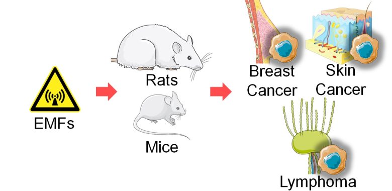
Next, I will present studies showing that exposure to EMFs increased breast cancer, skin cancer, and lymphoma in rats and mice.
Studies
Szmigielski et al. 1982
Female mice susceptible to breast cancer aged 6 weeks, equivalent to children/adolescents, were exposed to 2.45 GHz RF-EMF at a whole-body SAR of 2-3 W/kg or 6-8 W/kg for 2 hours per day for 10 months.
As a result, the onset of breast cancer was accelerated as the SAR value increased.
Acceleration of
Breast Cancer Onset
The larger the SAR value, the earlier the onset of breast cancer.
Also, male mice aged 6 weeks, equivalent to children/adolescents, were painted on their skin with benzopyrene, a carcinogen, and then exposed to 2.45 GHz RF-EMFs at a whole body SAR of 2-3 W/kg or 6-8 W/kg for 2 hours per day for 10 months.
As a result, the onset of skin cancer was accelerated as the SAR value increased.
Acceleration of
Skin Cancer Onset 1
The larger the SAR value, the earlier the onset of skin cancer.
Also, male mice aged 6 weeks, equivalent to children/adolescents, were exposed to 2.45 GHz RF-EMFs at a whole-body SAR of 2-3 W/kg for 2 hours per day for 1 month or 3 months.
Next, benzopyrene, a carcinogen, was painted on their skin and followed up.
As a result, the onset of skin cancer was accelerated as the duration of prior exposure to EMFs increased.
Acceleration of
Skin Cancer Onset 2
The longer the duration of prior exposure to EMFs, the earlier the onset of skin cancer.
Chou et al. 1992
Male rats aged 8 weeks, equivalent to adolescents, were exposed to pulse-modulated 2.45 GHz RF-EMFs at a whole-body SAR of 0.15-0.4 W/kg for 21.5 hours per day for 25 months.
As a result, primary cancers increased.
Increase in Primary Cancers
The odds of primary cancers increased 4-fold with the EMF exposure.
Based on the data described in the paper, I calculated the degree of increase for metastatic cancers.
An increase in metastatic cancers was observed in the exposed group, and it was especially more pronounced for lymphomas.
Increase in Metastatic Cancers
The odds of metastatic cancer increased 3-fold with the EMF exposure, and 10-fold for lymphomas.
