Consequences of Cell Death

Up to this point, I have explained the pathways of cell death due to cell cycle arrest from DNA damage, due to mitochondrial damage, and due to neuroinflammation.
In turn, cell death affects health by leading to a decrease in sperm count, a decrease in follicle count, infertility, malformations, low-birth-weight babies, neurodegenerative diseases, a decline in memory, developmental disorders, arrhythmias, COPD, asthma, diabetes, and more.
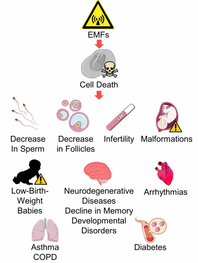
Table of ContentsAll_Pages
Male Infertility and a Decrease in Sperm Count
Cell Death in the Testes
Cell division actively takes place in the male testes, producing 50 to 100 million sperm per day.
The following is the process of sperm formation in the testis.
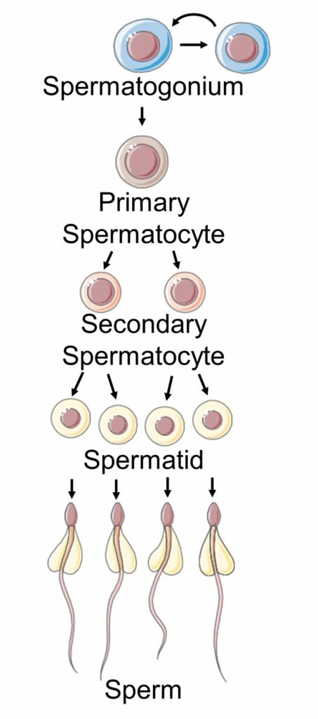
First, spermatogonia, stem cells in the testis, divide to form primary spermatocytes. Spermatogonia also divide and renew themselves.
Primary spermatocytes divide to form secondary spermatocytes, and further secondary spermatocytes divide to form spermatids.
When the spermatids have grown, they turn into sperm.
As described above, since cell division actively takes place in the testes, when DNA damage and replication stress occur due to oxidative stress and the cell cycle was arrested, it is likely that a large amount of cell death occurs, resulting in a decrease in sperm count.
In fact, experiments using rats or mice have confirmed that exposure to EMFs from cell phones caused cell cycle arrest of testicular cells and induced cell death. (Kesari et al. 2011, Pandey et al. 2016, Bin-Meferij and El-Kott 2015)
And as high as 90% of male infertility problems are related to count (Kumar and Singh 2015), thus, decreased sperm count is directly related to male infertility.
Therefore, it follows that an increase in cell death in the testes due to cell cycle arrest leads to a decrease in sperm, and an increase in male infertility.

Male Infertility and a Decrease in Sperm due to EMFs
And in fact, EMF exposure has been shown to damage the testes, decrease sperm count, decrease sperm quality, and increase male infertility. These studies can be found on page 1 of the following article.
Reproductive Abnormalities (Page 1)
Damage to the Testes Male Infertility and a Decrease in Sperm

It has been shown that exposure to EMFs decreases sperm count, increases male infertility, female infertility, miscarriage, birth defects, and low-birth-weight babies, and decreases the male birth rate. In recent years, sperm count is on the decline, infertility, miscarriage, birth defects, low-birth-weight babies, and preterm … Read the Full Article
Female Infertility
Cell Death in the Ovaries
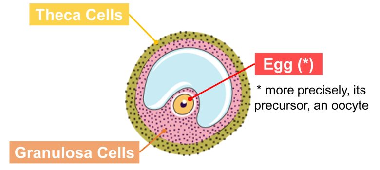
Within the ovary are numerous groupings of cells called follicles, each containing a single egg (or more precisely, its precursor, an oocyte). The follicle is composed of an egg, granulosa cells, which suport the egg, and theca cells, which are the envelope that covers the granulosa cells.
The following is the process of follicle maturation.
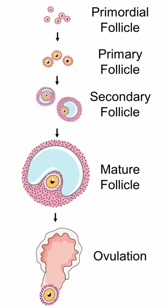
Follicles are normally stored in the ovary in a dormant state; these are called primordial follicles.
Several of these primordial follicles are stimulated by hormones to awaken and grow in stages over about a year to primary follicles, secondary follicles, and mature follicles.
When a follicle reaches a mature follicle, it ovulates.
Follicles that do not reach ovulation during this process lead to the death of the follicle, called follicular atresia.
This follicular atresia, however, has been demonstrated to be caused by cell death of the granulosa cells within the follicle. (SUGINO 2005)

Follicular atresia has been shown to be strongly associated with female infertility (Yao et al. 2021), and a study found a 3-fold increase in cell death of granulosa cells in women with unexplained infertility (İdil et al. 2004).
Therefore, it follows that an increase in cell death in the ovaries leads to an increase in the number of follicular atresia and an increase in female infertility.

Female Infertility due to EMFs 1
In fact, EMF exposure has been shown to damage the ovaries, increase cell death of follicle granulosa cells, increase the number of follicular atresia, and increase female infertility. These studies can be found on page 1 of the following article.
Reproductive Abnormalities (Page 1)
Damage to the Ovaries Female Infertility

It has been shown that exposure to EMFs decreases sperm count, increases male infertility, female infertility, miscarriage, birth defects, and low-birth-weight babies, and decreases the male birth rate. In recent years, sperm count is on the decline, infertility, miscarriage, birth defects, low-birth-weight babies, and preterm … Read the Full Article
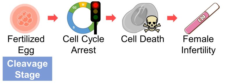
Cell Death of Fertilized Eggs
Once an egg is fertilized, the process from the fertilized egg to the fetus (pregnancy week 8), called embryogenesis, begins.
Embryogenesis is divided into the following stages: the cleavage stage, the blastulation, the gastrulation, and the organogenesis.

Cleavage Stage
During the cleavage stage, a fertilized egg undergoes multiple rounds of cell division, and the number of cells increases from 1 to 2 to 4 to 8 to 16, and so on, in powers of 2. Since division is without growth, individual cells become smaller as cleavage proceeds.

At this stage, cleavage of a fertilized egg may be arrested, which is a known cause for infertility. (Mohebi and Ghafouri-Fard 2019)
In fact, the arrested fertilized egg is heading toward cell death, and as many as 90% of fertilized eggs arrested in the cleavage stage were found to show signs of cell death. (Hardy 1999)
Cell death of fertilized eggs directly leads to loss of the embryo, resulting in implantation failure.
Therefore, it follows that an increase in cell death of fertilized eggs in the cleavage stage due to cell cycle arrest leads to an increase in female infertility.
Blastulation
As the number of cells increases during the cleavage stage, the cells of the fertilized egg align on the inner surface of the egg during the subsequent blastulation and form a fluid-filled cavity called a blastocoel. They also form an inner cell mass, an aggregation of cells that will later become the fetus.
This embryo, which forms 5-6 days after fertilization, is called a blastocyst (a blastula in non-mammals).

Pregnancy is regarded as established when the blastocyst implants in the placenta.
At the cleavage stage, embryos of good morphology show no sign of cell death. On the other hand, at the blastulation, scattered cell death is a common feature. (Hardy 1999)
However, there appears to be a correlation between increased cell death and decreased embryo quality; in blastocysts with good morphology, cell death was less than 10%, but in blastocysts with poor morphology, cell death was as high as 27%. (Hardy 1999)
And it has been shown that implantation rates of high quality blastocysts are high and implantation rates of low quality blastocysts are low. (Balaban et al. 2000)
In mammals including humans, failure in implantation leads to early embryonic loss and infertility. (Seshagiri et al. 2009)
Therefore, it follows that an increase in cell death of blastocysts in the blastulation due to cell cycle arrest, etc., leads to an increase in female infertility.

Female Infertility due to EMFs 2
And in fact, EMF exposure has been shown to damage blastocysts, increase their cell death, and increase female infertility. These studies can be found on page 1 of the following article.
Reproductive Abnormalities (Page 1)
Damage to Fertilized Eggs Female Infertility

It has been shown that exposure to EMFs decreases sperm count, increases male infertility, female infertility, miscarriage, birth defects, and low-birth-weight babies, and decreases the male birth rate. In recent years, sperm count is on the decline, infertility, miscarriage, birth defects, low-birth-weight babies, and preterm … Read the Full Article
Malformations
Cell Death in Embryos
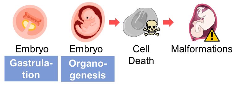
Cell death during embryogenesis can cause malformations.
During the gastrulation, exposure to alcohol, the acne medication isotretinoin, the rheumatoid medication methotrexate, hypoxia, ionizing radiation, and hyperthermic stress can cause craniofacial malformations. A common feature for many is excessive cell death for which the embryo may be unable to compensate. (Sulik et al. 1988)
During the gastrulation and organogenesis, exposure to thalidomide generates oxidative stress in the early developing limbs, and induces cell death, resulting in limb truncations. (Knobloch and Rüther 2008)
During organogenesis, exposure to nicotine generates oxidative stress in the neural tube, and induce cell death, resulting in neural tube defects such as spina bifida. (Zhao and Reece 2005)
Therefore, it follows that an increase in cell death of embryos in the gastrulation and organogenesis leads to an increase in malformations.
Malformations by EMFs
And in fact, EMF exposure has been shown to cause embryo malformations and increase birth defects. These studies can be found on page 1 of the following article.
Reproductive Abnormalities (Page 1)

It has been shown that exposure to EMFs decreases sperm count, increases male infertility, female infertility, miscarriage, birth defects, and low-birth-weight babies, and decreases the male birth rate. In recent years, sperm count is on the decline, infertility, miscarriage, birth defects, low-birth-weight babies, and preterm … Read the Full Article
Low-Birth-Weight Babies
Cell Death in the Placenta
The fetus receives oxygen and nutrients from the maternal blood via tissues called villi in the placenta.
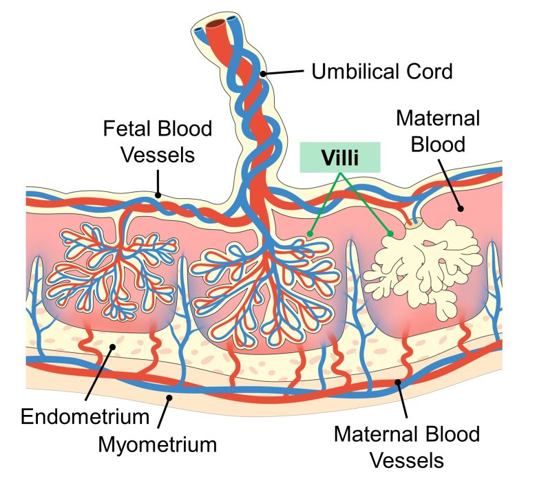
Maldevelopment of villi are characteristic of the placenta in fetal growth restriction, resulting in insufficient oxygen and nutrient supply for the fetus. (Scifres and Nelson 2009)
An increase in cell death of villi has been shown in fetal growth restriction, and it is thought to be the cause of maldevelopment of villi. (Scifres and Nelson 2009)
Therefore, it follows that an increase in cell death of placental villi leads to an increase in fetal growth restriction and an increase in low-birth-weight babies.

Cell Death Throughout the Body
Cell death in other tissues should also lead to fetal growth restriction, although there is little evidence in the literature.
It is likely that EMFs can ignore the placental barrier and interfere directly with cells throughout the fetal body.
It is not difficult to imagine that if a fetus, who is in the process of undergoing rapid cell division, is exposed to EMFs, ROS increase in cells throughout the body, which causes cell cycle arrest, leading to an increase in cell death and resulting in fetal growth restriction.
For example, in an experiment using mice, excessive generation of ROS through genetic manipulation resulted in low-birth-weight fetuses and delayed neonatal development. (Ishii et al. 2011)
Therefore, it is conceivable that an increase in cell death in the fetal body due to cell cycle arrest leads to an increase in fetal growth restriction and an increase in low-birth-weight babies.

Low-Birth-Weight Babies
And in fact, EMF exposure has been shown to delay growth and increase low-birth-weight babies. These studies can be found on page 1 of the following article.
Reproductive Abnormalities (Page 1)
Delayed Growth Low-Birth-Weight Babies

It has been shown that exposure to EMFs decreases sperm count, increases male infertility, female infertility, miscarriage, birth defects, and low-birth-weight babies, and decreases the male birth rate. In recent years, sperm count is on the decline, infertility, miscarriage, birth defects, low-birth-weight babies, and preterm … Read the Full Article
Neurodegenerative Diseases
Neurodegeneration from Brain Immune Responses
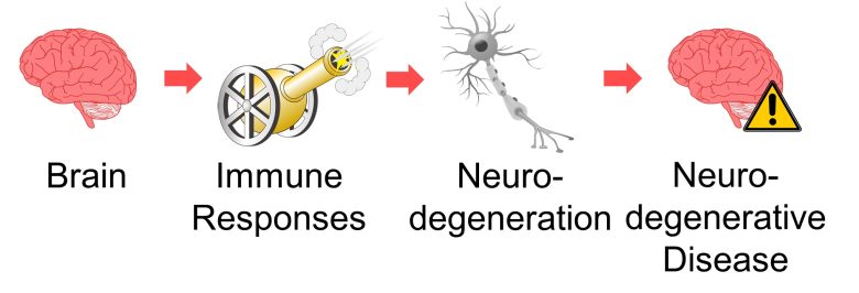
Glial cells such as microglia and astrocytes are activated as immune responses in the brain.
Activated astrocytes (reactive astrocytes) are increased, especially in Alzheimer's disease and ALS (Ben Haim et al. 2015), and have been implicated in the onset and progression of several neurodegenerative diseases (Phatnani and Maniatis 2015).
Chronic microglial activation may also contribute to the development and progression of neurodegenerative disorders. (Smith et al. 2012)
In addition, as mentioned earlier, brain immune responses causes excitotoxicity.
Excitotoxicity is known to be observed in numerous neurodegenerative diseases, including Huntington's disease, Alzheimer's disease, ALS, Parkinson's disease, and multiple sclerosis. (Salińska et al. 2005)
Therefore, it can be said that immune reactions in the brain lead to neurodegenerative diseases.
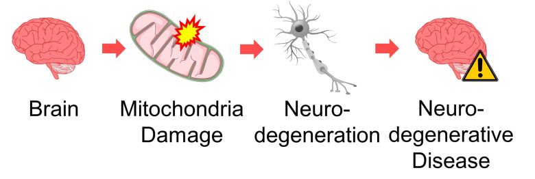
Neurodegeneration from Mitochondrial Damage
Mitochondria may have an important role in ageing-related neurodegenerative disorders like Parkinson's disease, Alzheimer's disease, Huntington's disease and ALS. (Federico et al. 2012)
Oxidative stress and structurally and functionally damaged mitochondria are early and prominent features of Alzheimer's disease. And a vicious downward spiral involving ROS and mitochondrial deficits is critical to pathogenesis of the disease. (Wang et al. 2014)
It has also been observed that the opening of the mitochondrial permeability transition pore caused excitotoxicity in neurons, resulting in cell death. (Liu et al. 2015, Choi et al. 2013)
Therefore, it can be said that mitochondrial damage in the brain leads to neurodegenerative diseases.
Neurodegenerative Diseases by EMFs
And in fact, EMF exposure has been shown to damage the brain and increase neurodegenerative diseases, especially Alzheimer's disease and ALS. These studies can be found on page 2 of the following article.
Brain Abnormalities (Page 2)
Damage to the Brains Neurodegenerative Diseases

It has been shown that exposure to EMFs increases behavior problems, ADHD, neurodegenerative diseases such as Alzheimer's and ALS, depression, suicide, electromagnetic hypersensitivity, biological clock disturbance, and neurotransmitter dysfunction. In recent years, academic performance is on the decline, and developmental disorders such as ADHD and … Read the Full Article
Decline in Memory
Hippocampal Atrophy
The hippocampus is a part of the brain involved in memory, and in various pathologies where hippocampal atrophy (reduction in volume) has been observed, declines in memory have also been observed.
The pathology of Alzheimer's disease begins with hippocampal atrophy (Dhikav and Anand 2011), and a decline in memory is among the first symptoms (Jahn 2013).
Patients with major depression has reduced hippocampus (Videbech 2004), and show a decline in memory (Burt et al. 1995).
Studies have shown that low-birth-weight infants have reduced hippocampus and show a decline in memory. (Isaacs et al. 2000, Aanes et al. 2019)
Studies have shown that in patients with brain traumatic brain injury have reduced hippocampus and show a decline in momory (Bigler et al. 1996, Monti et al. 2013)
Neurodegeneration results in neuronal damage and cell death, which leads to atrophy of brain tissue.
Therefore, it is conceivable that neurodegeneration in the brain leads to atrophy of hippocampus, which in turn leads to decline in memory.

Decline in Memory by EMFs
And in fact, EMF exposure has been shown to degenerate neurons and decrease their number in the hippocampus and other parts, and decline memory. These studies can be found on page 2 of the following article.
Brain Abnormalities (Page 2)
Damage to the Brain Decline in Memory

It has been shown that exposure to EMFs increases behavior problems, ADHD, neurodegenerative diseases such as Alzheimer's and ALS, depression, suicide, electromagnetic hypersensitivity, biological clock disturbance, and neurotransmitter dysfunction. In recent years, academic performance is on the decline, and developmental disorders such as ADHD and … Read the Full Article
Developmental Disorders
Brain Atrophy
There is now conclusive evidence that children with ADHD are associated with globally decreased brain volumes relative to control children. (Krain and Castellanos 2006)
Recently, a cross-sectional mega-analysis found amygdala, accumbens, and hippocampus reduction in ADHD. (Hoogman et al. 2017)
Marked Purkinje cell loss is the most consistent finding in the autistic disorder, and there is a theory that the selective vulnerability of the Purkinje cell may play a role in the etiology of autism. (Kinnear Kern 2003)
Neurodegeneration results in neuronal damage and cell death, which leads to atrophy of brain tissue.
Therefore, it is conceivable that neurodegeneration in the brain leads to atrophy of brain tissue, which in turn leads to developmental disorders such as ADHD and autism.

Developmental Disorders by EMFs
And in fact, EMF exposure has been shown to degenerate hippocampal neurons and Purkinje cells and decrease their number, and increase hyperactivity and behavior problems and ADHD. These studies can be found on page 2 of the following article.
Brain Abnormalities (Page 2)
Damage to the Brain Hyperactivity Behavior Problems and ADHD

It has been shown that exposure to EMFs increases behavior problems, ADHD, neurodegenerative diseases such as Alzheimer's and ALS, depression, suicide, electromagnetic hypersensitivity, biological clock disturbance, and neurotransmitter dysfunction. In recent years, academic performance is on the decline, and developmental disorders such as ADHD and … Read the Full Article
Arrhythmias
Cell Death and Fibrosis in the Heart Muscle
The loss of functional cells such as heart muscle cells and their replacement by structural component such as collagen is called fibrosis (scarring). (Piek et al. 2016)
Heart fibrosis results in stiffening and remodeling (structual and functional change) of the heart, and is increasingly recognized as a major cause of death in many chronic heart diseases (Piek et al. 2016)
For example, fibrosis is an important risk factor for arrhythmias. (Kazbanov et al. 2016)
Bradycardia, an arrhythmia where pulse slows down, results from conduction block in the sinus node or atrioventricular node, which is often caused by fibrosis. (Verheule and Schotten 2021)
The sinus node is the heart muscle that produces the heart's rhythm and is known as the natural pacemaker.
It have been demonstrated that increased fibrosis in ventricles, chambers that pump blood to the lungs and the body, promotes abnormal heart's electrical activity that can initiate ventricular fibrillation, which is the most lethal arrhythmia. (Morita et al. 2014)
Therefore, it follows that cell death in the heart causes fibrosis, which in turn leads to an increase in arrhythmias and other cardiac diseases.

Arrhythmias by EMFs
And in fact, EMF exposure has been shown to increase cell death and fibrosis in the heart muscle including the sinus node, and increase arrhythmias, particularly bradycardia. These studies can be found on page 3 of the following article.
Various Organs Abnormalities (Page 4)
Damage to the Heart Arrhythmias

It has been shown that exposure to EMFs increases arrhythmias, heart attacks (myocardial infarctions), COPD, asthma, diabetes, autonomic imbalance, impaired liver and kidney function, and cataracts. In recent years, arrhythmia, COPD, asthma, diabetes, liver disease, kidney disease, and cataracts are on the rise, and EMFs … Read the Full Article
COPD and Asthma
Chronic Inflammation
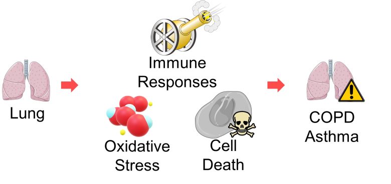
Chronic obstructive pulmonary disease (COPD) is characterised by chronic inflammation of the airways and progressive destruction of alveoli (air sacs), a process that in most cases is initiated by cigarette smoking. (Demedts et al. 2006)
Several mechanisms are involved in the development of the disease: infiltration of inflammatory cells into the lung (immune responses), imbalance of protein degradation, oxidative stress, and cell death. (Demedts et al. 2006)
EMFs are involved in immune responses, oxidative stress, and cell death among these, as explained up to this point.
Asthma is also characterized by chronic inflammation of the airways with infiltration of inflammatory cells into the lung (Hamid and Tulic 2009), and is associated with a strong oxidative stress that is a result of both increased oxidant forces and decreased antioxidant capacity. (Sahiner et al. 2011).
Therefore, immune response, oxidative stress, and cell death in the lungs may lead to an increase in COPD and asthma.
Impaired Lung Function in Fetushood
COPD is conventionally thought of as a disease of adult smokers. However, there is important epidemiological evidence that early life events, including pre-birth influences on lung growth, program the child to be at increased risk for future COPD. (Bush 2008)
A study result suggested that airway function throughout adult life may be determined during fetal development and the first few months of post-birth life. (Gibson and Simpson 2009)
Also, a study found that children developing asthma by age 7 had a lung function deficit and increased airway sensitivity as newborns and that this lung function deficit progressed to age 7. (Bisgaard et al. 2012)
Therefore, cell death in the fetal lungs may lead to impaired lung function, leading to an increase in asthma in childhood and COPD in adulthood.

COPD and Asthma by EMFs
And in fact, EMF exposure has been shown to increase oxidative stress in the lungs, and increase COPD and asthma.
Also, there is a study showing that EMFs impairs lung growth in fetuses.
These studies can be found on page 3 of the following article.
Various Organs Abnormalities (Page 4)
Damage to the Lungs COPD and Asthma

It has been shown that exposure to EMFs increases arrhythmias, heart attacks (myocardial infarctions), COPD, asthma, diabetes, autonomic imbalance, impaired liver and kidney function, and cataracts. In recent years, arrhythmia, COPD, asthma, diabetes, liver disease, kidney disease, and cataracts are on the rise, and EMFs … Read the Full Article
Diabetes
Cell Death in the Pancreas
Beta cells in the islets of Langerhans within the pancreas secrete insulin, a hormone that lowers blood glucose levels.
In type 1 diabetes, β-cell mass is reduced by as much as 70–80% at the time of diagnosis, In type 2, 25-50%. (Bucchieri et al. 2002)
A decrease in beta cells directly leads to a decrease in insulin secretion, which increases blood glucose levels and leads to diabetes.
Therefore, it follows that an increase in cell death in the islets of Langerhans leads to decreases in beta cell mass and insulin secretion, resulting in an increase in blood glucose and diabetes.

Diabetes by EMFs
And in fact, EMF exposure has been shown to damage the islets of Langerhans in the pancreas, decrease their cell volume, decrease insulin secretion, increase blood glucose levels, and increase diabetes. These studies can be found on page 3 of the following article.
Various Organs Abnormalities (Page 4)
Damage to the Pancreas Diabetes

It has been shown that exposure to EMFs increases arrhythmias, heart attacks (myocardial infarctions), COPD, asthma, diabetes, autonomic imbalance, impaired liver and kidney function, and cataracts. In recent years, arrhythmia, COPD, asthma, diabetes, liver disease, kidney disease, and cataracts are on the rise, and EMFs … Read the Full Article

