Consequences of Mutations

Up to this point, I have explained the pathways of mutations from DNA damage.
In turn, mutations affect health by leading to infertility, miscarriage, genetic disorders, cancer, and more.
There are also mechanisms that detect large-scale mutations and induce cell death.
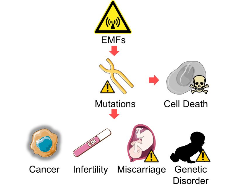
Table of ContentsAll_Pages
Cell Death
Chromosome Structural Abnormalities and Aneuploidy
First, large-scale mutations, such as dicentric and acentric chromosomes, unrepaired chromosome breaks, and aneuploidy, are induced to cell death by the tumor suppressor gene p53, etc. (Schwartz 1997, Belloni et al. 2008, Karlseder et al. 1999, Lips and Kaina 2001, Li et al. 2010, Thompson and Compton 2010)
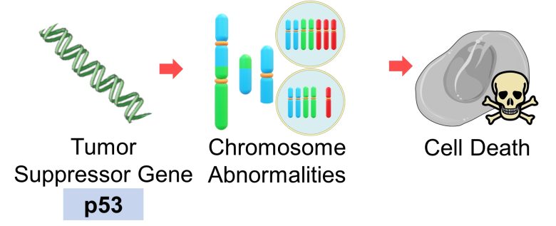
However, this mechanism for detecting chromosome abnormalities is not perfect.
For example, it is known that aneuploidy remain undetected in eggs (oocytes). This is because either deterioration of the detection mechanism due to aging, or eggs does not have an inherent mechanism to detect certain types of segregation defects. (Jones 2007)
Also, if the gene that constitutes this detection mechanism itself becomes dysfunctional due to mutations, the mutated cells will remain in place, opening the way for cancer, etc.
Miscarriage
Chromosome Aneuploidy
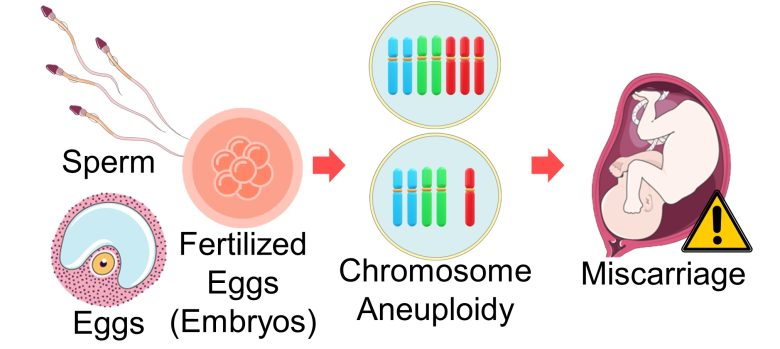
Chromosome aneuploidy is the most primary factor causing miscarriage. (Hassold and Hunt 2001)
In particular, among aneuploidies, monosomy, which is missing one chromosome, results in miscarriage for all but chromosome 21. (Fisher et al. 2012)
Most aneuploidy is derived from eggs, but some is also derived from sperm. (Nagaoka et al. 2012)
For example, the rate of sperm chromosome aneuploidy in recurrent miscarriage group was twice as high as fertile control group. (Carrell 2003)
Therefore, it follows that miscarriage will increase when chromosome aneuploidy occurs during the formation of eggs and sperm or during the growth of fertilized eggs (embryos).
Chromosome Structural Abnormalities
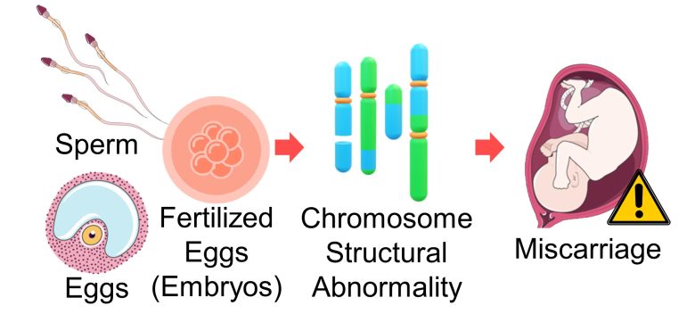
As mentioned above, the primary cause of miscarriage is aneuploidy, but some also result from structural abnormalities.
50% of miscarriages are due to chromosome abnormalities, of which as many as 86% are chromosome aneuploidies, while 6% are structural chromosome abnormalities and 8% are other genetic mechanisms. (Goddijn and Leschot 2000)
Therefore, it can be said that chromosome structural abnormalities can also increase miscarriage.
Miscarriage by EMFs
And in fact, EMF exposure has been shown to increase miscarriage and decrease the number of births. These studies can be found on page 1 of the following article.
Reproductive Abnormalities (Page 1)
Miscarriage Decrease in Births

It has been shown that exposure to EMFs decreases sperm count, increases male infertility, female infertility, miscarriage, birth defects, and low-birth-weight babies, and decreases the male birth rate. In recent years, sperm count is on the decline, infertility, miscarriage, birth defects, low-birth-weight babies, and preterm… Read the Full Article
Infertility
Chromosome Aneuploidy
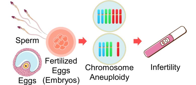
Chromosome aneuploidy is not only a factor in miscarriage, but also in infertility.
Aneuploidy rates found in first trimester pregnancy are much lower than those found in eggs and embryos before implantation. This suggests that a considerable proportion of embryos with chromosome abnomalities is lost before recognition. (Munné et al. 2003)
That is, as aneuploidy increases, the number of embryos lost before implantation increases, so pregnancy does not occur, thus, female infertility increases.
Also, infertility men have higher levels of sperm aneuploidy and the incidence of aneuploidy increases proportionally with the severity of the infertility. (Templado et al. 2013)
Therefore, it follows that infertility will increase when chromosomal aneuploidy occurs during the formation of eggs and sperm or during the growth of fertilized eggs (embryos).
Infertility by EMFs
And in fact, EMF exposure has been shown to increase male and female infertility. These studies can be found on page 1 of the following article.
Reproductive Abnormalities (Page 1)
Male Infertility Female Infertility

It has been shown that exposure to EMFs decreases sperm count, increases male infertility, female infertility, miscarriage, birth defects, and low-birth-weight babies, and decreases the male birth rate. In recent years, sperm count is on the decline, infertility, miscarriage, birth defects, low-birth-weight babies, and preterm… Read the Full Article
Genetic Disorders
Chromosome Aneuploidy
Chromosome aneuploidy is the most primary factor causing congenital defects. (Nagaoka et al. 2012)
For example, it is well known that trisomy of chromosome 21 causes Down syndrome and trisomy of chromosome 18 causes Edwards syndrome.
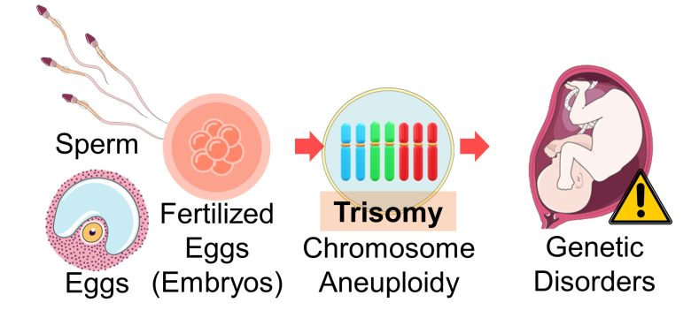
However, most other chromosome trisomies and all monosomies are fatal to the embryos and result in miscarriage.
Chromosome Structural Abnormalities
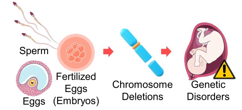
When chromosome deletions occur during the formation of eggs and sperm or during the growth of fertilized eggs (embryos), genetic disorders called deletion syndromes, which are associated with malformations and mental retardation, will develop.
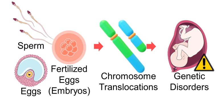
Translocations, on the other hand, are typically benign, but some can cause genetic disorders such as Down syndrome. (Wilch and Morton 2018)
Birth Defects by EMFs
And in fact, EMF exposure has been shown to increase birth defects, including genetic disorders. These studies can be found on page 1 of the following article.
Reproductive Abnormalities (Page 1)

It has been shown that exposure to EMFs decreases sperm count, increases male infertility, female infertility, miscarriage, birth defects, and low-birth-weight babies, and decreases the male birth rate. In recent years, sperm count is on the decline, infertility, miscarriage, birth defects, low-birth-weight babies, and preterm… Read the Full Article
Cancer
Base Substitution
Tumor Suppressor Gene p53
When a mutation occurs in DNA, it changes the nature of the protein produced by the DNA.
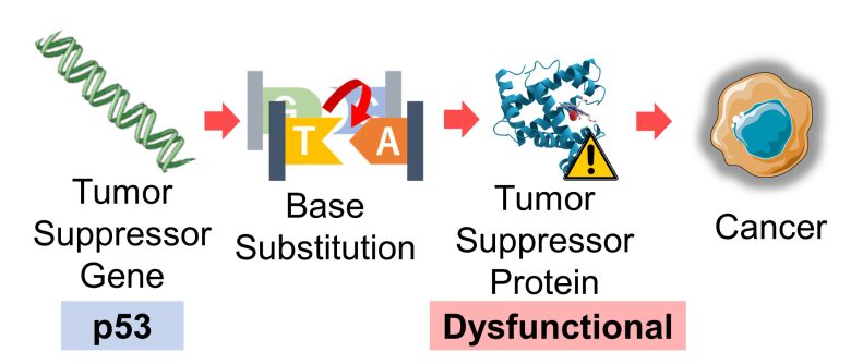
In particular, it is well known that mutations in tumor suppressor genes make their proteins dysfunctional, leading to cancer.
For example, in 50% of human cancers, the protein of the tumor suppressor gene p53 is inactivated. As many as 80% of these cases are due to base substitution (missense) mutations. (Soussi and Lozano 2005)
Proto-Oncogene Ras
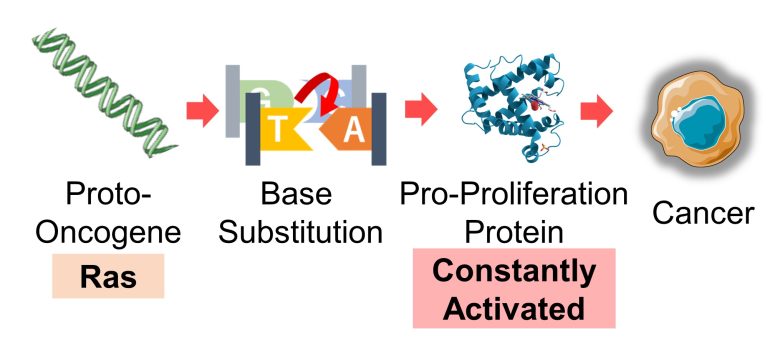
It is also well known that mutations in genes that trigger cell proliferation, called proto-oncogenes (*), make their proteins constantly activated, leading to cancer.
Mutated proto-oncogenes are referred to as oncogenes.
For example, the proto-oncogene Ras acquires cancer-causing potential when mutated by base substitution, which is observed in 20% of human cancers. (Prior et al. 2020)
Base Pair Deletions
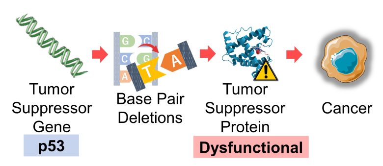
A correlation between base pair deletions and cancer has also been shown, although not as strong as with base substitutions.
Tumor Suppressor Gene p53
Of the 740 mutations in the cancer suppressor gene p53 collected from human cancers, base pair deletions and insertions were observed in 10% of them. (Jego et al. 1993)
Chromosome Structural Abnormalities
Chromosome structural abnormalities can also bring about cancer.
Dicentric Chromosomes
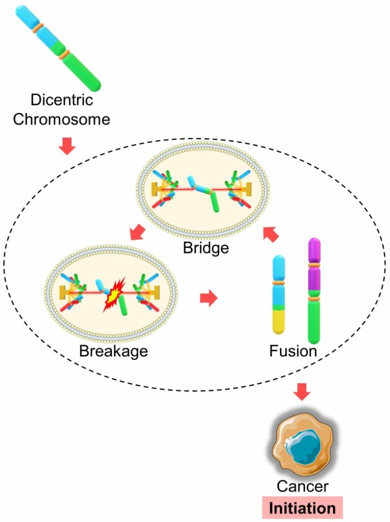
As mentioned earlier, dicentric chromosomes trigger cascading chromosomal damage called the "breakage–fusion–bridge cycle," which is strongly associated with cancer initiation. (Gascoigne and Cheeseman 2013)
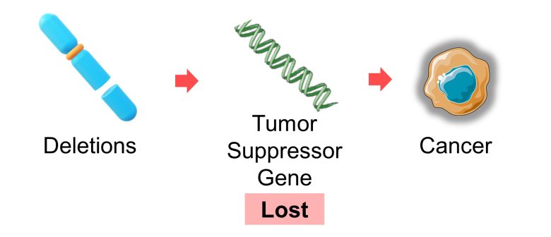
Deletions
Deletions ofen result in loss of a tumor suppressor gene, leading to cancer. (Rabbitts 1994)
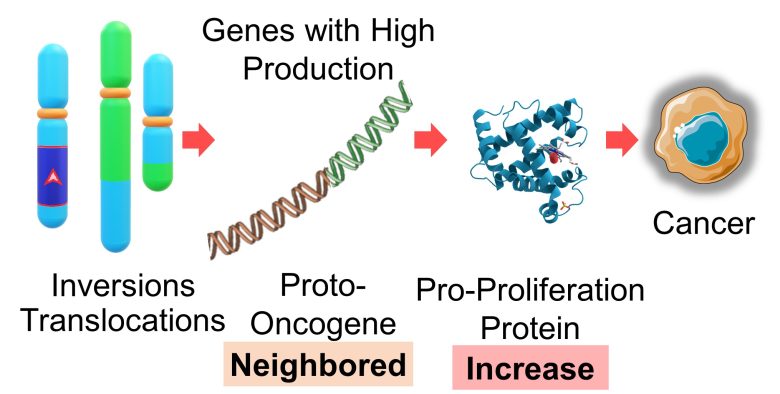
Inversions and Translocations
Inversions and translocations place proto-oncogenes near a gene with high production, such as antibodies, resulting in an abnormal increase in the production of proteins that promote proliferation, which in turn leads to cancer. (Rabbitts 1994, Nussenzweig and Nussenzweig 2010)
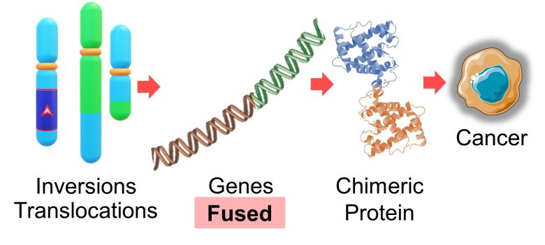
Aslo, inversions and translocations cause genes to fuse together, resulting in the production of proteins specific to cancer, called chimeric proteins, which also lead to cancer. (Rabbitts 1994, Nussenzweig and Nussenzweig 2010)
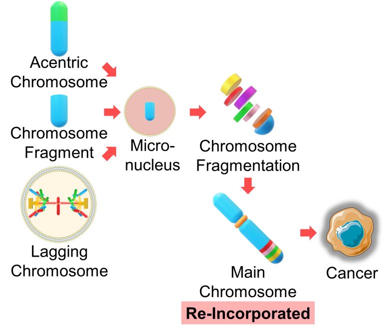
Acentric Chromosomes, Chromosome Fragments, Lagging Chromosomes
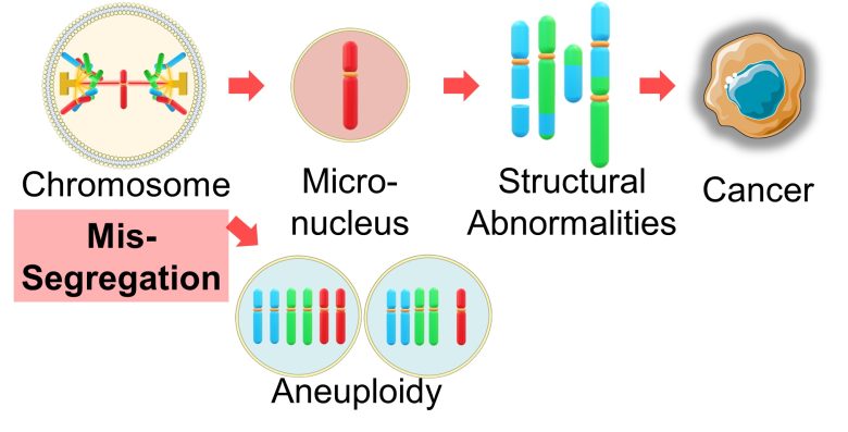
Chromosomal damage and formation of micronucleus are believed to play a significant role in the onset of many cancers. (Bhatia and Kumar 2012)
As mentioned earlier, it has been shown that the chromosome in the micronucleus undergo fragmentation and re-incorported into the main chromosomes, exacerbating chromosome structural abnormalities. (Crasta et al. 2012)
Therefore, micronuclei arising from acentric chromosomes, chromosome fragments, and lagging chromosomes can lead to cancer.
Chromosome Aneuploidy
It is not well understood whether chromosome aneuploidy causes cancer.
Micronuclei
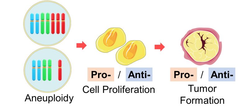
First, up to 90% of solid tumors are aneuploid (ranging from 26% to 99% across tumor types). (Ben-David and Amon 2019)
However, whether aneuploidy promotes tumor formation depends on the cellular context, and aneuploidy has been shown to both promote and suppress cell proliferation. (Ben-David and Amon 2019)
In addition, a 2019 US-UK study found that single-chromosome mis-segregation into a micronucleus are sufficient to cause various chromosome structual abnomalities (chromosome rearrangement), such as translocations, deletions, inversions, fragmentation, and more. (Ly et al. 2019)
That is, micronuclei arising simultaneously with aneuploidy can lead to cancer.
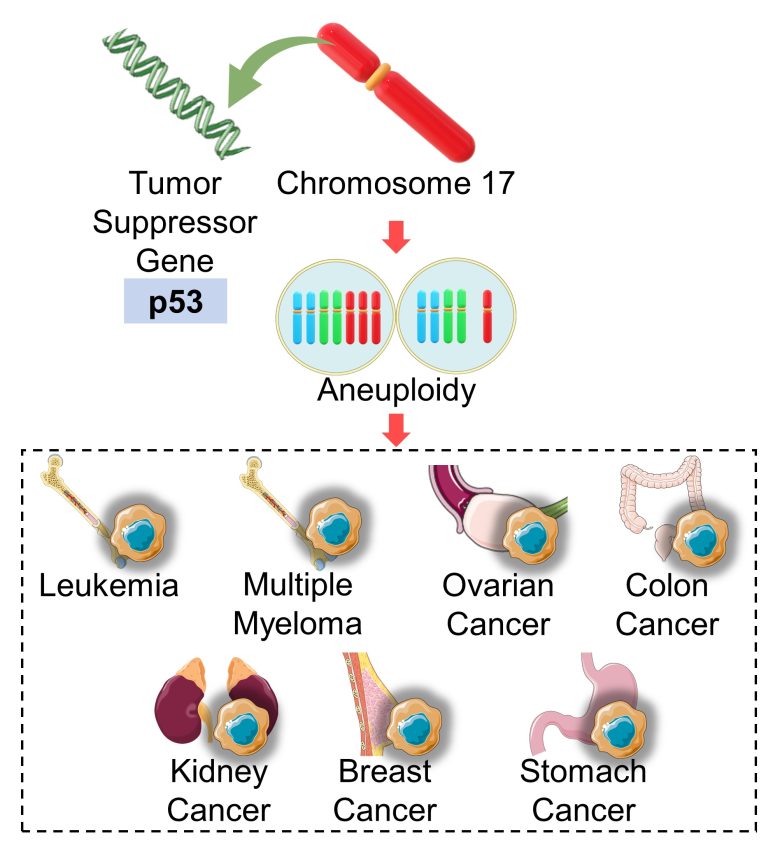
Chromosome 17
Apart from that, aneuploidy of chromosome 17, where the tumor suppressor gene p53 resides, is known to be observed in many cancers, including leukemia, multiple myeloma, ovarian cancer, colon cancer, kidney cancer, breast cancer, and stomach cancer. (Othman et al. 2001)
On the previous page, I presented studies showing increased aneuploidy in chromosome 17 due to EMF exposure.
Cancer by EMFs
And in fact, EMFs has been shown to increase wide-ranging cancers, including leukemia, lymphoma, brain tumors, breast cancer, and testicular cancer. These studies can be found on page 4 of the following article.
Cancer (Page 3)
Leukemia and Lymphoma Brain Tumors Breast Cancer Testicular Cancer Other Cancers
It has been shown that EMF exposure increases various types of cancer, including leukemia (especially lymphoid leukemia), lymphoma, breast cancer, testicular cancer, lung cancer, and pancreatic cancer. In recent years, these cancers are on the rise, and EMFs are likely to be one of the… Read the Full Article

