
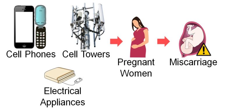
In this day and age, you might come off as a bit insane if you say that EMFs (Electric and Magnetic Fields) are bad for you.
But this is in fact what numerous scientific papers are pointing to.
I too was skeptical until I actually saw it with my own eyes.
In this article, I will present a total of 200 studies showing that EMFs from cell phones and other sources have adverse health effects.
The health effects shown in humans are extremely wide-ranging, as follows:
Reproductive Abnormalities
Male infertility, a decrease in sperm count, female infertility, miscarriage, birth defects, low-birth-weight babies, a decrease in male births, etc.
Brain Abnormalities
Behavior problems, ADHD, a decline in memory, dementia, Alzheimer's disease, ALS, electromagnetic hypersensitivity, depression, suicide, biological clock disturbance, neurotransmitter dysfunction, etc.
Cancer
Leukemia, lymphoma, brain tumors, breast cancer, testicular cancer, pancreatic cancer, lung cancer, etc.
Various Organ Abnormalities
Arrhythmias, heart attacks (myocardial infarctions), COPD, asthma, diabetes, autonomic imbalance, impaired liver and kidney function, cataracts, etc.
It has also been shown, mainly through animal experiments, that EMFs can actually damage various organs.
The damaged parts are also wide-ranging, including the testes, ovaries, fertilized eggs (embryos), brain, heart, lungs, pancreas, liver, and kidneys.
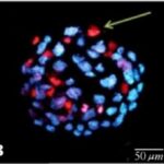
For example, below are the pictures taken from animal experiments, which show visible organ damage.
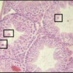
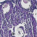
Damage to the Brain

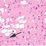
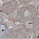
Due to exposure to cell phone EMFs, numerous vacuoles (cavities) occurred in the cerebral cortex of a brain. (Akakin et al. 2020)
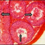
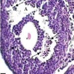
Damage to the Testes
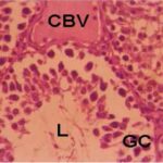
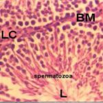
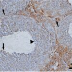
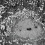
Due to exposure to smartphone EMFs, seminiferous tubules within a testis were damaged and sperm decreased. (Hasan et al. 2021)
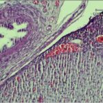
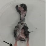
Damage to Fertilized Eggs (Embryos)

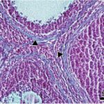
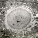
Due to EMF exposure, fertilized eggs (embryos) were considerably delayed in development and malformed. (Delgado et al. 1982)
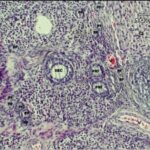
Damage to the Pancreas
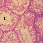
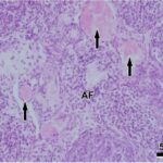
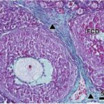
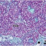
Due to cell phone exposure, most of the cells in the islet of Langerhans within a pancreas were dissociated. (Mortazavi et al. 2016)
These animal experiments back up the wide-ranging effects of EMFs on human health.
Chapters
It is also now getting clearer that EMFs affect health mainly by increasing reactive oxygen species. The mechanism is explained in the following article.
Mechanisms of How EMFs Affect Health

In the previous article, I presented numerous studies showing that EMFs (Electric and Magnetic Fields) have adverse health effects. This article will continue with an explanation of how this works. EMFs have been shown to cause adverse health effects, primarily by increasing reactive oxygen species… Read the Full Article
Abbreviations : RF-EMFs, Radio Frequency EMFs; ELF-EMFs, Extremely Low Frequency EMFs
Reproductive Abnormalities

It has been shown that exposure to EMFs decreases sperm count, increases male infertility, female infertility, miscarriage, birth defects, and low-birth-weight babies, and decreases the male birth rate.
In recent years, sperm count is on the decline, infertility, miscarriage, birth defects, low-birth-weight babies, and preterm births are on the rise, and the male birth rate is on the decline.
Many of these abnormalities began to increase around the time commercial cell phone service began, and EMFs are likely to be the major factor in their increase.
Recent Trends
Sperm Count :
All Continents
Infertility :
US
Japan
Birth Defects :
Down Syndrome
Edwards Syndrome
Gastroschisis
Cystic Kidney
Diaphragmatic Hernia
Hypospadias
Renal Agenesis
Miscarriage :
US
Sweeden
Low-Birth-Weight Babies :
US
Japan
Preterm Births :
US
Japan
World
Male Birth Rate :
US
Japan
Table of ContentsAll_Pages
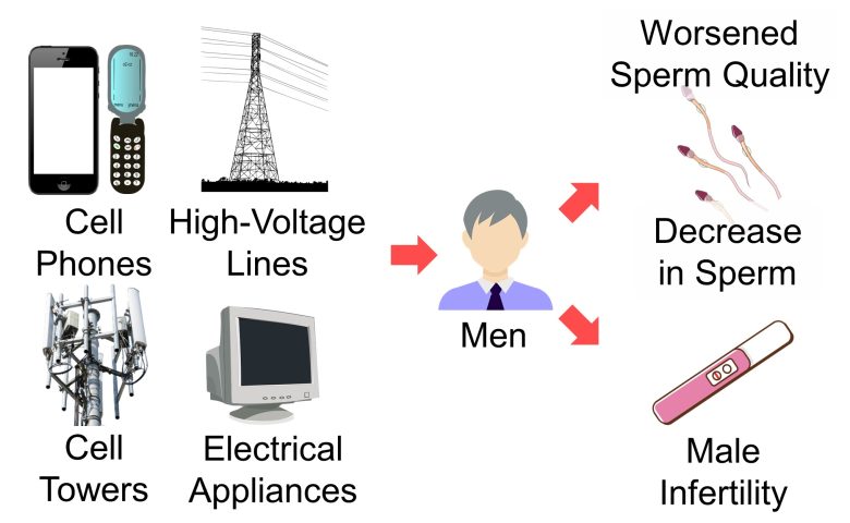
Male Infertility and a Decrease in Sperm Count
First, I will present studies showing that sperm count decreased, sperm quality worsened, and male infertility increased with exposure of men to EMFs from cell phones, cell towers, high voltage lines, and electrical appliances.
Stuties
Agarwal et al. 2008
For men aged on average 31 years attending an infertility clinic in Cleveland, Ohio, sperm count decreased, sperm motility and viability decreased, and sperm abnormalities increased as the time they spent per day on their cell phones increased.
Decrease in Sperm Count
The sperm count decreased by 40% with the cell phone use for 4 hours or more per day.
Decrease in Motility and Viability
Increase in Sperm Abnormalities
The percentage of normal-shaped sperm decreased by 50% with the cell phone use for 4 hours or more per day.
Ding et al. 2018
For men attending a genetics clinic in Shaanxi, China, reactive oxygen species (ROS) in their semen increased, sperm count decreased, sperm motility decreased, and DNA breaks in their sperm increased as the time they were exposed per day to the EMFs increased, emitted from smartphones, cell towers, and Wi-Fi.
Increase in ROS
Decreases in total antioxidant capacity and antioxidant activity mean an increase in ROS.
Decrease in Sperm Count
The sperm count decreased by 70% with the EMF exposure for more than 2 hours per day, emitted from cell phones, cell towers, and Wi-Fi.
Decrease in Motility
The comet assay is used to detect DNA breaks. In the comet assay, the more the DNA breaks, the more the tail DNA, the less the head DNA, and the larger the olive tail moment.
Increase in DNA Breaks
Kilgallon and Simmons 2005
For men aged 18-35 years recruited at the University of Western Australia, sperm count decreased, and sperm motility decreased when they carried their cell phones near their testes.
Decrease in Sperm Count
The sperm count decreased by 10% with the cell phones in the hip pockets or on the belts.
Decrease in Motility
The sperm motility decreased by 10% with the cell phones in the hip pockets or on the belts.
Fejes et al. 2005
For men aged on average 31 years undergoing fertility treatment at the University of Szeged in Hungary, sperm count decreased when they carried their cell phones near their bodies for a prolonged period.
Decrease in Sperm Count
The sperm count decreased by 10% when the cell phone was carried within 20 inches (50 cm) of the body for more than 20 hours.
El-Helaly et al. 2010
For men aged on average 30 years undergoing fertility treatment at the Mansoura University Hospital in Egypt, male infertility increased when they used video display terminals (VDTs) and computers (*) at workplaces.
When you use the wireless function of your computer, you are exposed to RF-EMFs.
Increase in Male Infertility
The odds of male infertility increased 8-fold with the use of VDTs and computers at workplaces.
Otitoloju et al. 2009
Male mice were kept for 6 months in a workplace complex with one cell tower in the neighborhood, or in a residential quarter with two cell towers.
As a result, sperm abnormalities increased as the number of cell towers increased in the neighborhood.
The strength of RF-EMFs was 0.001 μW/cm2 in the control facility, 0.063 μW/cm2 in the workplace complex, and 0.103 μW/cm2 in the residential quarter. (*)
It is calculated from the strength of the electric field described in the paper, as the impedance of free space 377 Ω.
Increase in Sperm Abnormalities
The sperm head abnormalities increased 19-fold with one cell tower in the neighborhood and 22-fold with two.
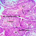
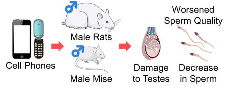
Damage to the Testes
Next, I will present studies showing that exposure to EMFs from cell phones and other sources damaged the testes of rats and mice, decreased sperm count, and worsened sperm quality.
Spermatogenesis and Infertility
A male testis contains numerous tubes called seminiferous tubules, where sperm are actively produced.
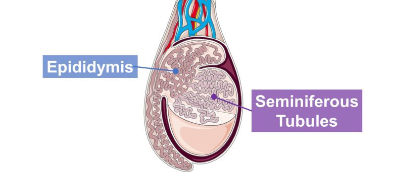
On the inner surface of the seminiferous tubule reside stem cells called spermatogonia. The spermatogonia divide and renew themselves, and some of them turn into spermatocytes. When the spermatocytes divide, they turn into spermatids. The spermatids then grow to become sperm.
The spermatogonium, spermatocyte, and spermatid are also called spermatogenic cells ( sperm-producing cells ).
These spermatogenic cells mature as they move inward into the center, where they become sperm and are transported to an epididymis, which stores sperm. Spermatogenic cells are supported and nourished by Sertoli cells, known as nurse cells.
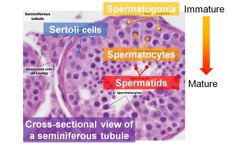
( Cited and modified from Health Jade )
And, it has been shown that exposure to EMFs damages seminiferous tubules in testes of rats and mice and decreases spermatogenic cells and sperm.
As high as 90% of male infertility problems are related to sperm count (Kumar and Singh 2015), so it can be said that decreased sperm count leads directly to male infertility.
Studies
Hasan et al. 2021
Smartphones were placed on the ceilings of breeding cages and kept on incoming calls for 40 or 60 minutes per day, and male mice aged 6 weeks, equivalent to children and adolescents, were exposed to their EMFs for 60 days.
As a result, the seminiferous tubules within the testes were damaged, and spermatogenic cells and sperm decreased.
A decrease in weight gain was also observed, which is described in the section on Delayed Growth.
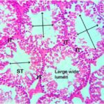
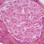
Damage to Seminiferous Tubules
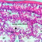
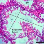
Due to the exposure to the smartphone EMFs, the seminiferous tubules were damaged, and spermatogenic cells were degenerated and decreased. In addition, the insides of the seminiferous tubules were hollowed out, and almost no sperm were present.
Bin-Meferij and El-Kott 2015
Test cell phones with a local SAR of 0.96 W/kg were placed in the center of breeding cages, and male rats aged 2 months, equivalent to adolescents, were exposed to their EMFs for 1 hour per day for 7 weeks.
As a result, reactive oxygen species (ROS) in blood increased, the seminiferous tubules were damaged, sperm count decreased, sperm motility and viability decreased, and sperm abnormalities increased.
Also, treatment with moringa, which has antioxidant properties, suppressed the damage above.
Increase in ROS
An increase in lipid peroxidation and a decrease in antioxidant activity mean an increase in ROS.
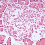
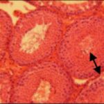
Damage to Seminiferous Tubules

Due to the exposure to the cell phone EMFs, the seminiferous tubules were damaged, spermatogenic cells were degenerated, and multinucleated giant cells (*) appeared. In addition, the insides of the seminiferous tubules were hollowed out, and almost no sperm were present. Treatment with the moringa, which has antioxidant properties, suppressed the damage.
Macrophages, immune cells, fuse with each other to form multinucleated giant cells, a hallmark of chronic inflammation. (McNally and Anderson 2011)
Decrease in Sperm Count
The sperm count decreased by 60% due to the exposure to the cell phone EMFs. Also, the moringa, which has antioxidant properties, suppressed it.
Increase in Sperm Abnormalities
Decrease in Viability and Motility
Pandey et al. 2016
Male mice aged 8-10 weeks, equivalent to adolescents, were exposed to RF-EMFs similar to those emitted from cell phones for 4 or 8 hours per day for 35 days.
As a result, the seminiferous tubules were damaged and atrophied, the testes shrank, sperm count decreased, sperm abnormalities increased, and DNA breaks in the testes increased as the exposure time per day to the RF-EMFs increased.
The damage above recovered to some extent as the time passed after the exposure.
Damage to Seminiferous Tubules

Due to the exposure to the RF-EMFs, immature spermatogenic cells sloughed off into the inside, decreasing sperm. In addition, multinucleated giant cells (*) appeared.
Macrophages, immune cells, fuse with each other to form multinucleated giant cells, a hallmark of chronic inflammation. (McNally and Anderson 2011)
Atrophy of Seminiferous Tubules
Shrinkage of Testes
Decrease in Sperm Count
Increase in Sperm Abnormalities
The comet assay is used to detect DNA breaks. In the comet assay, the more the DNA breaks, the more the tail DNA, the less the head DNA, and the larger the olive tail moment.
Increase in DNA Breaks
Kumar et al. 2014
Cell phones with a local SAR of 1.34 W/kg were placed and kept in talk mode for 2 hours per day, and male rats aged 10 weeks, equivalent to adolescents, were exposed to their EMFs at very close range for 60 days.
As a result, reactive oxygen species (ROS) in the sperm increased, sperm count decreased, the testes shrank, and DNA breaks in the sperm increased.
Increase in ROS
TBARS, a marker for lipid peroxidation, increased by 60% due to the exposure to the cell phone EMFs.
An Increase in lipid peroxidation means an increase in ROS.
Decrease in Sperm Count
The sperm count decreased by 20% due to the exposure to the cell phone EMFs.
Shrinkage of Testes
The comet assay is used to detect DNA breaks. In the comet assay, the more the DNA breaks, the further the tail migration, the longer the tail length, the more the %DNA in tail, and the larger the olive tail moment.
Increase DNA Breaks 1
In the comet assay, tail migration increased due to the exposure to the cell phone EMFs. This means an increase in DNA breaks.
Increase DNA Breaks 2
Female Infertility
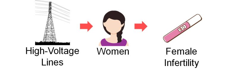
Next, I will present a study showing that female infertility increased with exposure of women to EMFs from high-voltage lines.
Studies
Esmailzadeh et al. 2019
For women aged on average 28 years attending a infertility clinic in Babol, Mazandaran, Iran, female infertility increased as the distance from their homes to high-voltage lines decreased.
Increase in Female Infertility
The odds of female infertility increased 4-fold within 550 yards (500 m) of the high-voltage lines.
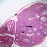
Damage to the Ovaries
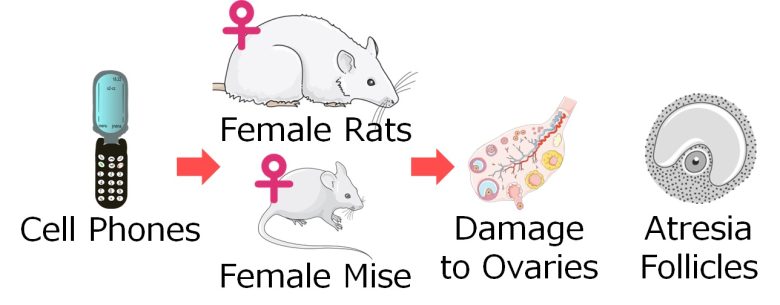
Next, I will present studies showing that exposure to EMFs from cell phones and other sources damaged the ovaries of rats and mice, decreased healthy follicle count, and increased atresia follicle count.
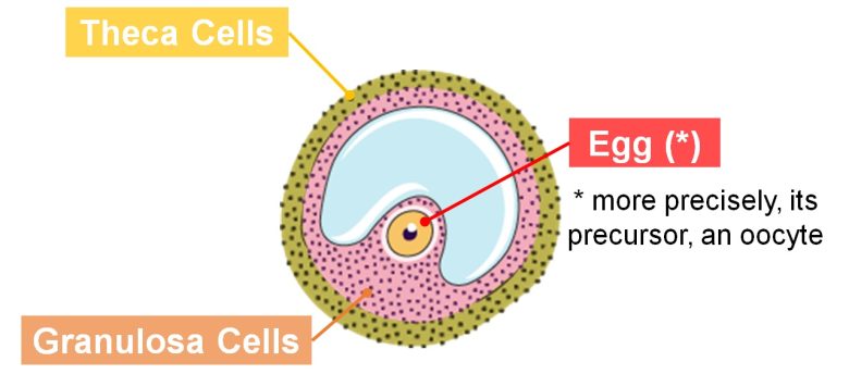
Folliculogenesis and Infertility
Within the ovary are numerous groupings of cells called follicles, each containing a single egg (or more precisely, its precursor, an oocyte). The follicle is composed of an egg, granulosa cells, which suport the egg, and theca cells, which are the envelope that covers the granulosa cells.
Follicles are normally stored in the ovary in a dormant state; these are called primordial follicles.
Several of these primordial follicles are stimulated by hormones to awaken and grow in stages over about a year to primary follicles, secondary follicles, and mature follicles.
When a follicle reaches the mature follicle, it ovulates.
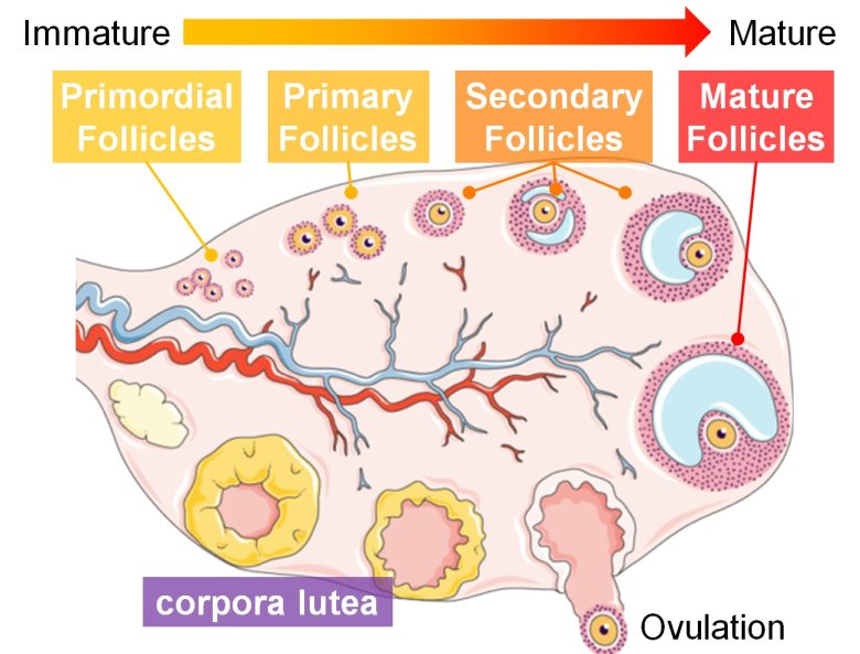
Follicles that do not reach ovulation during this process lead to the death of the follicle, called follicular atresia.
This follicular atresia, however, has been demonstrated to be caused by cell death of the granulosa cells within the follicle. (SUGINO 2005)
Follicular atresia has been shown to be strongly associated with female infertility (Yao et al. 2021), and a study found a 3-fold increase in cell death of granulosa cells in women with unexplained infertility (İdil et al. 2004).
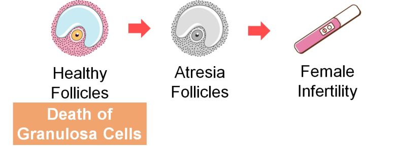
And, it has been shown that exposure to EMFs damages ovaries of rats and mice, increases cell death of granulosa cells in follicles, decreases primordial, secondary, primary, and mature follicle count, and increases atresia follicle count.
Studies
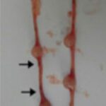
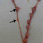
Türedi et al. 2016
Pregnant rats were exposed to 900 MHz RF-EMFs with a strength of 26.5 μW/cm2 for 1 hour per day for 1 week in late pregnancy.
As a result, in the born-daughter rats, the eggs in the primary follicles were lost, primordial, primary, and mature follicle count decreased, and atresia follicle count increased. The ovaries were damaged, and cell death of granulosa cells increased.
Loss of Eggs
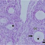
Due to the exposure to the RF-EMFs during pregnancy, eggs in the majority of primary follicles of the daughter rats were lost. Degeneration of granulosa cells and vascular congestion were also observed.
Decrease in Follicles
Increase in Atresia Follicles
The atresia follicle count of the daughter rats increased 2-fold due to the exposure to the RF-EMFs during pregnancy.
Increase in Cell Death 1
Due to the exposure to the RF-EMFs during pregnancy, cell death increased in the ovaries of the daughter rats, particularly of granulosa cells and theca cells.
Increase in Cell Death 2
The percentage of cell death increased 2-fold in the ovaries of the daughter rats due to the exposure to the RF-EMFs during pregnancy.
Degeneration of the Ovaries 1
Due to the exposure to the RF-EMFs during pregnancy, follicular cells degenerated and stromal tissue became fibrotic in the ovaries of the daughter rats.
Degeneration of the Ovaries 2
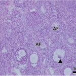
Rad et al. 2014
Pregnant mice were exposed to 50 Hz ELF-EMFs with a strength of 3 mT for 4 hours per day for 3 weeks throughout the entire pregnancy.
As a result, in the newborn daughter mice, the eggs degenerated.
In the grown-up daughter mice, primordial, primary, secondary, and mature follicle count decreased, and atresia follicle count increased. The ovaries were damaged, cell death of granulosa cells increased, and the oocytes degenerated.
Degeneration of Eggs in Newborns
Due to the exposure to the ELF-EMFs during pregnancy, eggs were degenerated in the ovaries of the newborn daughter mice.
Decrease in Follicles in Adults
Damage to Follicles in Adults
Due to the exposure to the ELF-EMFs during pregnancy, degeneration and cell death of granulosa cells were observed, and eggs degenerated in the ovaries of the grown-up daughter mice.
Decrease in Atresia Follicles in Adults
The percentage of atresia follicles increased 3-fold in the ovaries of the grown-up daughter mice due to the exposure to the ELF-EMFs during pregnancy.
Bakacak et al. 2015
Female rats aged 16 weeks, equivalent to adolescents, were exposed to 900 MHz RF-EMFs pulse-modulated at 217 Hz at a whole-body SAR of 0.018-4 W/kg for 2 hours per day for 35 days.
As a result, primordial follicle count decreased in the ovaries.
Decrease in Follicles 1
Due to the exposure to the RF-EMFs, primordial follicles decreased.
Decrease in Follicles 2
The primordial follicle count decreased by 50% due to the exposure to the RF-EMFs.
Panagopoulos et al. 2010
Female and male fruit flies, newly hatched from pupae, were placed in separate vials at a distance of 0 inch-39 inches (0 cm-100 cm) from a cell phone with a local SAR of 0.89 W/kg and exposed to its EMFs for 6 minutes per day for 6 days.
From the 3rd day of sexual maturity, females and males were placed in a vial together to mate and lay eggs.
As a result, cell death increased in the female's ovarioles (tubular components of insect ovaries) and the number of hatched pupae decreased as the distance to the cell phone increased.
Increase in Cell Death 1
The closer the distance to the cell phone, the more the cell death in the ovarioles.
Increase in Cell Death 2
The closer the distance to the cell phone, the higher the percentage of cell death in the ovarioles.
Decrease in Hatched Pupae
The closer the distance to the cell phone, the fewer the number of hatched pupae.
The decrease in the number of hatched pupae can be attributed to increased infertility in both males and females and decreased hatched rates due to the exposure to the cell phone EMFs.

Miscarriage
Next, I will present studies showing that miscarriage increased with exposure of pregnant women to EMFs from cell phones, cell towers, electrical appliances, and other sources.
Studies
Mahmoudabadi et al. 2015
For pregnant women aged 18-35 years in Tehran, Iran, compared to those with normal pregnancies, those with miscarriages had longer call time on a cell phone, had a higher percentage of app use, and had a higher percentage of carrying a cell phone close to the body.
Longer Call Time
The call time on a cell phone increased 3-fold for the women with miscarriages.
Higher App Use
The percentage of app use on a cell phone increased 2-fold for the women with miscarriages.
Carrying Closer to the Body
Based on the data described in the paper, I calculated the degree of increase for miscarriage.
Increase in Miscarriage
Using Apps
The odds of miscarriage increased 3-fold when they used apps on their cell phones during pregnancy.
Increase in Miscarriage
Carrying Near the Body
Zhou et al. 2015
For pregnat women aged 16-45 years seeking medical attention in Beijing Obstetrics and Gynecology Hospital, compared to those with normal pregnancies, those with miscarriages had a higher percentage of living near a cell tower.
Living Closer to Cell Tower
The percentage of living within 110 yards (100 m) of a cell tower increased 2-fold for the women with miscarriages.
Based on the data described in the paper, I calculated the degree of increase for miscarriage.
Increase in Miscarriage
Living Near Cell Towers
The risk of miscarriage increased 2-fold when they lived within 110 yards (100 m) of the cell towers during pregnancy.
Wang et al. 2013
For pregnant women in two towns in the Pearl-River Delta of China, miscarriage increased when the ELF-EMFs in the alleys in front of their houses were stronger.
Increase in Miscarriage
The hazard of miscarriage increased 1.7-fold with 0.1 µT or more of the ELF-EMFs in the alleys in front of the pregnant women's houses.
Li et al. 2017
For pregnant members of a major health maintenance organization (HMO), residing in the San Francisco Bay Area, miscarriage increased when the daily average ELF-EMFs they were exposed to were stronger.
Increase in Miscarriage
The hazard of miscarriage increased 2.7-fold with 0.25 μT or more of the average daily ELF-EMFs the pregnant women were exposed to.
The increase in hazard of miscarriage in the two studies above means a increase in miscarriage rates per unit of time (e.g., per week).
For your reference, I applied these hazard ratios of miscarriage to the 1981-1982 miscarriage rates by week in California (Goldhaber and Fireman 1991) to produce the following graph.
Increase in Miscarriage Rates
by Week
The miscarriage rate at 20 weeks of pregnancy increased 1.6-fold from 11% to 18% for 0.1 μT or more of ELF-EMFs and 2.5-fold to 27% for 0.25 μT or more.
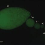
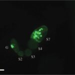
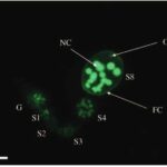
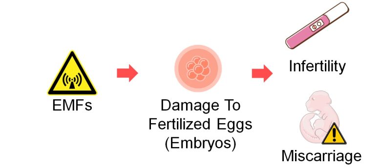
Damage to Fertilized Eggs (Embryos)
Next, I will present studies showing that exposure to EMFs damaged fertilized eggs (embryos), and increased infertility and miscarriage.
Embryogenesis
Once an egg is fertilized, the process from the fertilized egg to the fetus (pregnancy week 8), called embryogenesis, begins.
Embryogenesis is divided into the following stages: the cleavage stage, the blastulation, the gastrulation, and the organogenesis.
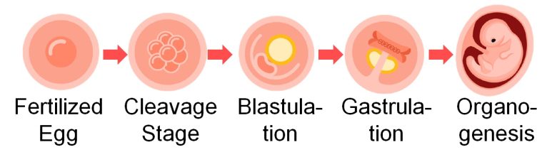
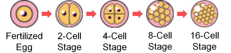
Cleavage Stage
During the cleavage stage, a fertilized egg undergoes multiple rounds of cell division, and the number of cells increases from 1 to 2 to 4 to 8 to 16, and so on, in powers of 2. Since division is without growth, individual cells become smaller as cleavage proceeds.
At this stage, cleavage of a fertilized egg may be arrested, which is a known cause for infertility. (Mohebi and Ghafouri-Fard 2019)
In fact, the arrested fertilized egg is heading toward cell death, and as many as 90% of fertilized eggs arrested in the cleavage stage were found to show signs of cell death. (Hardy 1999)
And, it has been shown that EMF exposure of mice and rats increases cell death in embryos, decreases embryo quality, increases embryonic death from the cleavage stage to the blastulation, and increases infertility.
Blastulation
As the number of cells increases during the cleavage stage, the cells of the fertilized egg align on the inner surface of the egg during the subsequent blastulation and form a fluid-filled cavity called a blastocoel. They also form an inner cell mass, an aggregation of cells that will later become the fetus.
This embryo, which forms 5-6 days after fertilization, is called a blastocyst (a blastula in non-mammals).

Pregnancy is regarded as established when the blastocyst implants in the placenta.
There appears to be a correlation between decreased blastocyst quality and increased cell death; in blastocysts with good morphology, cell death was less than 10%, but in blastocysts with poor morphology, cell death was as high as 27%. (Hardy 1999)
Implantation rates for high quality blastocysts are high and implantation rates for low quality blastocysts are low. (Balaban et al. 2000)
In mammals including humans, failure in implantation leads to early embryonic loss and infertility. (Seshagiri et al. 2009)
And, it has been shown that EMF exposure of mice and rats increases cell death in blastocysts, decreases embryo quality and increases embryonic death from the cleavage stage to the blastulation, decreases the number of implantations, and increases infertility.
Gastrulation and Later
Embryonic death after implantation is considered miscarriage.
The most primary factor causing miscarriage is chromosomal abnormalities. (Hassold and Hunt 2001)
Chromosomal abnormalities are also a factor in infertility and birth defects.
There are many experiments showing that EMF exposure of humans, animals, and plants can cause chromosome abnormalities. Details of these studies are presented in Mechanisms of How EMFs Affect Health.
Here, I will present a study showing that EMF exposure of rats increased pre-implantation or post-implantation embryo death and increased infertility or miscarriages.
Studies
Safian et al. 2016
Female mice were injected with ovulation-inducing drugs and allowed to mate with male mice, and the fertilized eggs (embryos) were removed the next day.
Next, cell phones with local SAR 0.683-0.725 W/kg were placed on top of the incubator and kept on talk mode for 30 minutes per day, and embryos on day 2 were exposed to their EMFs at a distance of 10 cm or further for 4 days.
As a result, the number of dead embryos increased from the cleavage stage to the blastulation, and embryo quality decreased. In addition, cell death increased in blastocysts on day 5.
Increase in Dead Embryos
Decrease in Embryo Quality
The percentage of high-quality embryos decreased from the cleavage stage to the blastulation due to the exposure to the cell phone EMFs.
Increase in Cell Death
in Blastocysts 1

Due to the exposure to the EMFs from the cell phones, cell death in the blastocysts increased and the quality of the blastocysts decreased.
Increase in Cell Death
in Blastocysts 2
Borhani et al. 2011
Female mice were exposed to 50 Hz ELF-EMFs with a strength of 500 μT for 4 hours per day for 12 days.
On the 8th day of exposure, they were injected with ovulation-inducing drugs and allowed to mate with males. This means embryos (fertilized eggs) were also exposed on and after the 8th day.
As a result, cell death of blastocysts increased, their number decreased, and infertility increased.
Increase in Cell Death in Blastocysts 1
Due to the exposure to the ELF-EMFs, cell death in blastocysts increased.
Increase in Cell Death in Blastocysts 2
The percentage of cell death in the blastocysts increased by 50% due to the exposure to the ELF-EMFs.
Decrease in Blastocysts
The number of blastocysts decreased by 40% due to the exposure to the ELF-EMFs. If it were human, it would directly lead to implantation failure, i.e., infertility.
Increase in Infertility
The percentage of pregnant mice decreased by 30% due to the exposure to the ELF-EMFs, indicating that the infertility increased.
Alchalabi et al. 2016
Female rats were allowed to mate with male rats, and fertilization was confirmed.
Next, the female rats were exposed to RF-EMFs similar to those from cell phones at a whole-body average SAR of 0.048 W/kg for either 1 or 2 hours per day.
The duration of exposure was either 1 week in early pregnancy, 2 weeks in early and mid-pregnancy, or 3 weeks throughout the entire pregnancy.
As a result, reactive oxygen species (ROS) in fetal blood increased and infertility/miscarriage increased as the time and duration they were exposed to the RF-EMFs increased.
Also, a decrease in the fetal weight, and malformations were observed.
Increase in ROS
An increase in lipid peroxidation and a decrease in antioxidant activity means an increase in ROS.
Increase in
Infertility/Miscarriage 1
Due to the exposure to the RF-EMFs for 2 weeks in early and mid-pregnancy, embryonic death increased at the uterine horns, indicating that infertility/miscarriage increased.
Increase in
Infertility/Miscarriage 2
Data from Week 1 of Pregnancy
Data from Week 2 of Pregnancy
Data from Week 3 of Pregnancy
Malformations
Partial malformations occurred due to the exposure to the RF-EMFs during pregnancy.
Decrease in Births
Next, I will present studies showing that the number of births decreased due to simultaneous exposure of males, females, and fertilized eggs (embryos) to EMFs from radio towers or cell towers.
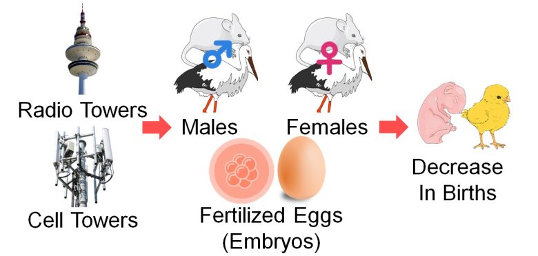
These studies will confirm the three combined factors of male and female infertility and increased miscarriage or decreased hatch rates due to exposure to EMFs.
I will also present a study showing that the number of pregnant females decreased due to either male or female EMF exposure in rat breeding pairs, i.e., both males and females became infertile.
Studies
Magras and Xenos 1997
On top of Mount Chortiati in Thessaloniki, Greece, was a dence area of radio towers called Antenna Park, where almost 100 commercial TV and FM-radio broadcasting transmitters were situated as of 1997.
This study was undertaken in response to residents' health concerns about Antenna Park.
Breeding pairs of mice aged 2 months were kept for 6 months in a village at the foot of the mountain and in a hillside near Antenna Park and were allowed to mate 5 times during this period.
The RF-EMFs' strength was 1.1 μW/cm2 in the village, 0.168 μW/cm2 in the hillside, and 0.0001 μW/cm2 at the laboratory that served as a control facility.
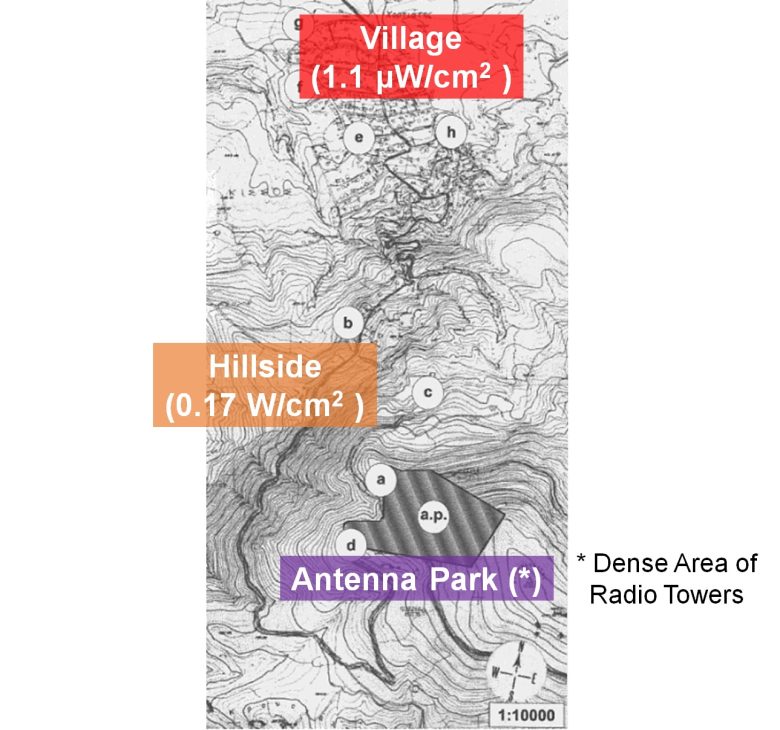
As a result, the number of births decreased as the exposure duration increased and as the strength of the EMFs increased, and eventually, no more pups were born.
Decrease in Births
The longer the exposure to the EMFs and the stronger the EMFs, the fewer the number of births. In the low-exposure hillside, the number of births was zero at the 5th mating and at the 3rd mating in the high-exposure village.
The decrease in births can be attributed to increased infertility in both males and females and increased miscarriage due to the exposure to the the radio tower EMFs.
Balmori 2005
For storks in Valladolid, Spain, a percentage of nests without chicks increased and the number of young birds that left the nest decreased when they nested near a cell tower.
The strength of RF-EMFs was 1.48 μW/cm2 within 220 yards (200 m) of the cell tower and 0.075 μW/cm2 over 300 m. (*)
It is calculated from the strength of the electric field described in the paper, as the impedance of free space 377 Ω.
Increase in Nests Without Chicks
The percentage of nests without chicks increased 12-fold within 220 yards (200 m) of the cell tower.
Birds That Left the Nests
As for the decrease in the number of young birds that left the nest, only a modest effect was observed when limited to the nests with chicks, suggesting that the cell tower EMFs mainly caused the increase in the percentage of nests without chicks.
The increase in the percentage of nests without chicks can be attributed to increased infertility in both males and females and decreased hatched rates due to the exposure to the cell tower EMFs.
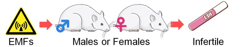
Al‐Akhras et al. 2001
Adult male and female rats were exposed to 50 Hz ELF-EMFs with a strength of 25 μT for 90 days.
After that, the rats were allowed to mate in combinations of the exposed males and the unexposed females, and of the unexposed males and the exposed females.
As a result, in the both combinations, percentages of pregnant females decreased, indicating that the both exposed males and females became infertile.
The first is the combination of the exposed males and the unexposed females.
Male Infertility
The percentage of pregnant females decreased by 50% due to the exposure of males to the ELF-EMFs, indicating that the males became infertile.
Increase in Fetus Resorption
The percentage of fetal resorption increased 9-fold due to the exposure of males to the ELF-EMFs, indicating that the sperm of the males deteriorated.
Avoiding EMF exposure restored the percentage of pregnant females, but there was no improvement in the percentage of fetal resorption. This may be because, for example, proliferation of sperm stem cells (spermatogonia) restored sperm count of the males, while mutated spermatogonia were not eradicated and remained.
The next is the combination of the unexposed males and the exposed females.
Female Infertility
The percentage of pregnant females decreased by 40% due to the exposure of females to the ELF-EMFs, indicating that the females became infertile.
Decrease in Implantations
The number of implantations decreased by 50% due to the exposure of females to the ELF-EMFs. If it were human, it would directly lead to implantation failure, i.e., infertility.
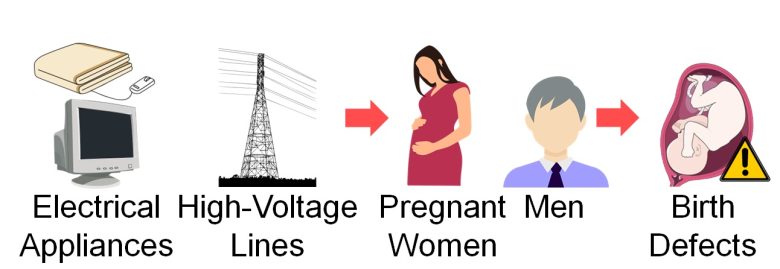
Birth Defects
Next, I will present studies showing that birth defects increased with exposure of pregnant women to EMFs from electrical appliances and other sources.
I will also present a study showing that birth defects in children increased with exposure of men to EMFs from high-voltage lines.
Studies
Ericson and Källén 1986
For pregnant employees throughout Sweden, birth defects in their babies increased when they had used video display terminals (VDTs) at work.
This was more pronounced as the time they spent per week on VDTs increased in early pregnancy.
Increase in Birth Defects
The odds of birth defects in the babies increased by 60% with the use of VDTs during pregnancy and 70% in early pregnancy.
Increase in Birth Defects
in Early Pregnancy
The odds of birth defects in the babies increased 2.0-fold with the use of VDTs for 10 hours or more per week in early pregnancy, and 2.3-fold for 20 hours or more.
Adjusted odds ratios are not provided in this paper.
Nordström et al. 1983
For male employees of the Swedish State Power Board and of one Swedish power company, congenital malformations in their children increased and also male infertility increased when they had worked at high-voltage substations.
A decrease in the male birth rate was also observed, which is described in the section on Decrease in the Male Birth Rate.
Increase in Congenital Malformations
The prevalence of congenital malformations in the children increased 6-fold with the work in the high-voltage substations.
Increase in Male Infertility
The prevalence of male infertility increased 3-fold with the work in the high-voltage substations.
Li et al. 1995
Among pregnant women in seven counties of western Washington State, for those with a history of subfertility (*), congenital urinary tract anomalies in their babies increased when they had used electric blankets.
a history of trying for more than 12 months without getting pregnant
This was more pronounced with early pregnancy use and as the use time increased.
Congenital Urinary Tract
Anomalies
Congenital anomalies of the kidney and urinary tract are the most common cause of prenatally diagnosed developmental malformations (Stonebrook et al. 2019). Cystic kidney and renal agenesis, which I mentioned at the beginning of this chapter as being on the rise in recent years, are included in this disease.
Becker and Becker 1986
At the time, Vernon Township in New Jersey had the world's highest civilian concentration of microwave sources with 3 earth stations, each with at least 3 satellite antennas and 29 terrestrial antennas within 5 miles of the center of town.
Residents began to notice an abnormally high incidence of health problems in some areas of the township, which led to the creation of a citizens' group that initiated an epidemiological study in the township by themselves.
They found that birth defects, malignancies, and chromosomal abnormalities were not randomly distributed throughout the town but occurred in clusters.
55% of the reported cases occurred in areas where only 27% of the residential population lived, and these clusters appeared to be associated with the projected beam paths from the antennas at the earth stations.
Among others, a higher number of cases of Down syndrome in Vernon Township was found compared to the national average.
Increase in Down Syndrome
The number of observed cases of Down syndrome was 4 times higher than the national average in Vernon Township, exposed to the microwaves from the earth stations.
The strength of the microwaves from the antenna at the earth station was as follows.
Microwave Strength
Even at 600 yards (550 m) from the antenna, the microwaves were quite strong, at 10 μW/cm2 or more.
See page 5 for dangerous levels of EMFs, including microwaves.
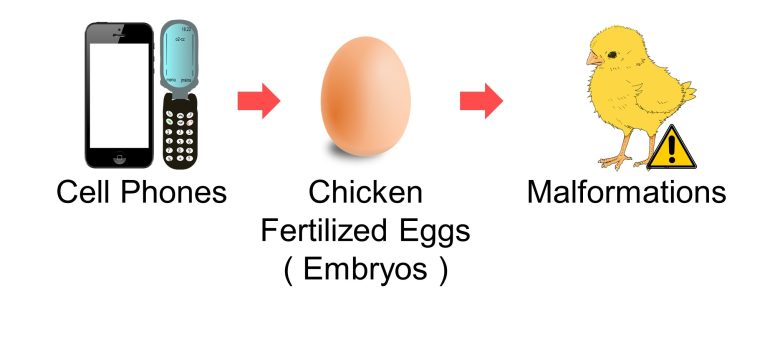
Malformations
Next, I will present studies showing that exposure to EMFs from cell phones and other sources increased malformations in chickens.
Studies
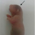
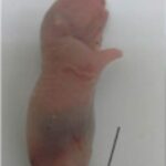
Delgado et al. 1982
Fertilized chicken eggs (embryos) were exposed to ELF-EMFs at frequencies of 10, 100, and 1000 Hz and with strengths of 0.1, 1, and 10 μT for 48 hours in nine combinations.
As a result, malformations in the embryos increased, and the most pronounced effect was produced with the 100 Hz x 1.2 μT combination from the morphological aspect.
This is similar to those emitted from high-voltage lines and electrical appliances, and pregnant women should especially be careful.
Increase in Malformations 1
Embryos were malformed due to the ELF-EMF exposure. Notably, there were considerable delays in development.
The developmental delays can be explained in terms of EMFs causing death to proliferating cells. For more information, see Mechanisms of How EMFs Affect Health.
Increase in Malformations 2
The malformation rate increased in various parts of the body due to the ELF-EMF exposure, especially in the nervous system, by a factor of 7-8.
Increase in Malformations 3
The combinations of 100Hz,1000Hz x 1.2μT had the most pronounced effect, with the 100% malformation rate.
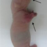
Augustianath et al. 2023
2G cell phones with a local SAR of 1.36 W/kg or 4G cell phones with a local SAR of 1.12 W/kg were placed on the ceilings of incubators and kept on incoming calls for 50 or 90 minutes per day, and fertilized chicken eggs (embryos) were exposed to their EMFs untile they hatched.
As a result, malformations in the embryos increased
In addition, the hatch rate decreased as the exposure time increased and as the SAR value increased (*).
However, since the communication protocols also differ, therefore the waveforms of the EMFs also differ, it cannot be simply a matter of SAR value.
Increase in Malformations
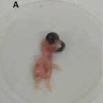

On day 10 of incubation, due to the exposure to the cell phone EMFs, the heads and legs of the chicks were malformed.

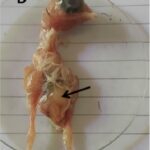
On day 15 of incubation, due to the exposure to the cell phone EMFs, the legs and the guts of the chicks were malformed.


On the 21th day when they hatched, due to the exposure to the cell phone EMFs, the legs of the chicks were malformed and they had difficulty standing straight.
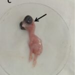
Decrease in Hatch Rates
The hatch rate decreased by 60% due to the exposure for 90 minutes per day from the 2G cell phones and by 40% from the 4G cell phones.
Un-Hatched Chick

Due to the exposure to the cell phone EMFs, one eye was missing, the beak was crossed, and the yolk sac didn't retract and remain in place.
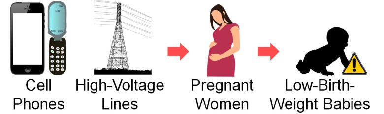
Low-Birth-Weight Babies
Next, I will present studies showing that low-birth-weight babies increased with exposure of pregnant women to EMFs from cell phones and high-voltage lines.
Low-birth-weight babies face health problems
A low-birth-weight baby is a baby who weighs less than 5.5 pounds (2,500 g) at birth; the standard birth weight is between 5.5 pounds and 8.8 pounds (2,500 g and 4,000 g).
In the U.S., about 1 in 12 babies is born with a low birth weight. (Cleveland Clinic)
Babies born with low birth weights have the following risks for a variety of health problems. (Cleveland Clinic)
Immediate Problems
Low oxygen levels at birth.
Infections.
Jaundice.
Breathing issues due to immature lungs.
Nervous system issues, including bleeding inside the brain.
Digestive system issues, including serious inflammation of the intestines.
Long-Term Progblems
Delayed motor and social development.
Learning differences.
Health conditions when they grow up, including high blood pressure, heart disease, obesity, and diabetes.
And it has been shown that low-birth-weight babies increase with EMF exposure.
Studies
de Vocht et al. 2014
For pregnant women in Northwest England, newborn birth weight decreased, and low-birth-weight babies increased as the distance from their homes to high-voltage lines or substations increased.
Also, an increase in spontaneous preterm births was observed.
Decrease in Birth Weight
The newborn birth weight decreased by 7.48 ounces (212 g) when the pregnant women's homes were within 55 yards (50 m) of the high-voltage lines or the substations.
Increase in Low-Birth-Weight Baby
The odds of low-birth-weight babies increased 3-fold when the pregnant women's homes were within 55 yards (50 m) of the high-voltage lines or the substations.
Increase in Spontaneous Preterm Births
The odds of spontaneous preterm births increased 2-fold when the pregnant women's homes were within 55 yards (50 m) of the high-voltage lines or the substations.
Lu et al. 2017
For pregnant women in Kumamoto, Japan, newborn birth weight decreased and also infant emergency transport increased when they had used their cell phones excessively.
A decrease in the male birth rate was also observed, which is described in the section on Decrease in the Male Birth Rate.
Decrease in Birth Weight
The newborn birth weight decreased by 4.55 ounces (129 g) with the pregnant women's excessive use of their cell phones.
Increase in Emergency Transport
The odds of infant emergency transport increased 8-fold with the pregnant women's excessive use of their cell phones.
Boileau et al. 2020
For pregnant women in Haute-Vienne, France, fetal growth restriction (fetal poor growth) increased when they spent more time per day on their cell phones.
Fetal growth restriction is commonly defined using small for gestational age (SGA) birth (in bottom 10% for birth weight) as a proxy. (Hutcheon et al. 2021)
Increase in Fetal Growth Restriction
The odds of fetal growth restriction increased by 50% when the pregnant women used their cell phones for 30 minutes or more per day.
No strong correlation was found, but this may be related to the fact that cell phone use for more than 4 hours per day was excluded as "aberrant" without any particular explanation.
Delayed Growth
Next, I will present studies showing that exposure to EMFs from smartphones and other sources delayed the growth of fetuses and pups of rats and mice.

Studies
Cao et al. 2006
Pregnant mice were exposed to 50 Hz ELF-EMFs with a strength of 1.2 mT for 8 hours per day for 3 weeks throughout the entire pregnancy.
As a result, the number of births decreased, fetal weight gain decreased, and growth after birth was delayed.
Decrease in Births
The number of births decreased by 40% due to the EMF exposure during pregnancy, indicating that miscarriage increased.
Decrease in Weight Gain
The fetal weight gain decreased by 40% due to the EMF exposure during pregnancy.
Delayed Growth
Eye opening and teething were delayed in the pups born due to the EMF exposure during pregnancy, indicating that growth after birth was delayed.
Other observations included malformations in the fetuses in the group exposed to the EMF.
Hasan et al. 2021
Smartphones were placed on the ceilings of breeding cages and kept on incoming calls for 40 or 60 minutes per day, and male mice aged 6 weeks, equivalent to children and adolescents, were exposed to their EMFs for 60 days.
As a result, weight gain decreased.
Damage to testes was also observed, which is described in the section on Damage to Testes.
Decrease in Weight Gain
The rate of weight gain in the unexposed group was 60%, while the rate of weight gain in the exposed group was 10%, a drastic decrease in the rate of weight gain.
Sangun et al. 2014
Pregnant rats were exposed to 2450 MHz RF-EMF for 1 hour per day at a whole-body average SAR of 0.143 W/kg for 3 weeks throughout the entire pregnancy, and then the born-daughter rats were exposed from three weeks of age to the beginning of puberty (equivalent to childhood).
As a result, reactive oxygen species (ROS) in the brain and ovaries increased, hormone imbalance occured, and the onset of puberty was delayed.
Increase in ROS
An increase in oxidative stress means an increase in ROS.
Hormone Imbalance
Delayed Onset of Puberty
The onset of estrus (heat) was delayed by 20% due to the exposure to the EMFs during childhood and by 30% during the fetushood and childhood.
Also, food and water intake increased, but conversely, weight gain decreased.
Increase in Food
and Water Intake
Decrease in Weight Gain
The weight gain didn't change due to the exposure to the EMFs during childhood and decreased by 20% during the fetushood and childhood.
No change or a decrease in weight gain despite an increase in food and water intake suggests that some abnormalities in the metabolic system occurred.
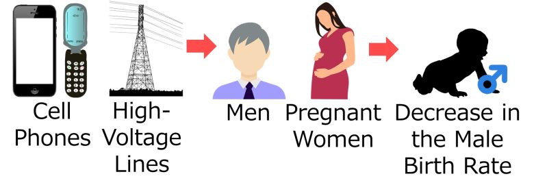
Decrease in the Male Birth Rate
Next, I will present studies showing that the male birth rate decreased with exposure of men to EMFs from high-voltage lines.
I will also present a study showing that the male birth rate decreased with exposure of pregnant women to EMFs from cell phones.
Influence of Enviramental and Occupational Exposure of Parents
More than 100 studies have examined whether environmental or occupational exposures of parents affect the sex ratio of their offspring at birth. (Terrell et al. 2011)
Regarding paternal exposure, the evidence is mounting that dioxins reduce the male birth rate. (Terrell et al. 2011)
There is also some evidence that paternal exposure to dibromochloropropane (soil fumigant), lead, methylmercury, non-ionizing radiation (=EMFs), ionizing radiation treatment for childhood cancer, boron, or g-force reduces the male birth rate. (Terrell et al. 2011)
On the other hand, few studies have found higher or lower sex ratios associated with maternal exposures. (Terrell et al. 2011)
Studies in humans and animals have found a reduction in the number of male births associated with lower male fertility, but the mechanism by which environmental hazards might change the sex ratio has not yet been established. (Terrell et al. 2011)
As we have seen up to this point, many studies have shown that EMFs lower male fertility. It would therefore not be surprising if EMFs were also an environmental hazard that reduces the male birth rate.
Studies
Knave et al. 1979
For male employees of the Swedish State Power Board and of one Swedish power company, the number of births, especially boy births, decreased when they had worked at high-voltage substations.
Decrease in Births
Nordström et al. 1983
For male employees of the Swedish State Power Board and of one Swedish power company, the male birth rate decreased when they had worked at high-voltage substations.
An increase in congenital malformations in their children and male infertility was also observed, which is described in the section on Birth Defects.
Decrease in the Male Birth Rate
The male birth rate decreased with the work at the high-voltage substations.
Lu et al. 2017
For pregnant women in Kumamoto, Japan, the male birth rate decreased when they had used their cell phones excessively.
A decrease in newborn birth weight was also observed, which is described in the section on Low-Birth-Weight Babies.
Decrease in the Male Birth Rate
The male birth rate decreased with the pregnant women's excessive use of their cell phones.